 Movie
Movie Controller


+ Open data

- Basic information
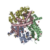
| Entry | Database: PDB / ID: 6o84 | |||||||||||||||||||||||||||||||||
|---|---|---|---|---|---|---|---|---|---|---|---|---|---|---|---|---|---|---|---|---|---|---|---|---|---|---|---|---|---|---|---|---|---|---|
| Title | Cryo-EM structure of OTOP3 from xenopus tropicalis | |||||||||||||||||||||||||||||||||
 Components Components | LOC100127796 protein,LOC100127796 protein,OTOP3,LOC100127796 protein,LOC100127796 protein | |||||||||||||||||||||||||||||||||
 Keywords Keywords | MEMBRANE PROTEIN / Otopetrin / proton / channel | |||||||||||||||||||||||||||||||||
| Function / homology | Otopetrin / Otopetrin / proton channel activity / proton transmembrane transport / identical protein binding / plasma membrane / Proton channel OTOP3 Function and homology information Function and homology information | |||||||||||||||||||||||||||||||||
| Biological species | ||||||||||||||||||||||||||||||||||
| Method | ELECTRON MICROSCOPY / single particle reconstruction / cryo EM / Resolution: 3.92 Å | |||||||||||||||||||||||||||||||||
 Authors Authors | Chen, Q.F. / Bai, X.C. / Jiang, Y.X. | |||||||||||||||||||||||||||||||||
| Funding support |  United States, 5items United States, 5items
| |||||||||||||||||||||||||||||||||
 Citation Citation | Journal: Elife / Year: 2019 Title: Structural and functional characterization of an otopetrin family proton channel. Authors: Qingfeng Chen / Weizhong Zeng / Ji She / Xiao-Chen Bai / Youxing Jiang / Abstract: The otopetrin (OTOP) proteins were recently characterized as proton channels. Here we present the cryo-EM structure of OTOP3 from (XtOTOP3) along with functional characterization of the channel. ...The otopetrin (OTOP) proteins were recently characterized as proton channels. Here we present the cryo-EM structure of OTOP3 from (XtOTOP3) along with functional characterization of the channel. XtOTOP3 forms a homodimer with each subunit containing 12 transmembrane helices that can be divided into two structurally homologous halves; each half assembles as an α-helical barrel that could potentially serve as a proton conduction pore. Both pores open from the extracellular half before becoming occluded at a central constriction point consisting of three highly conserved residues - Gln-Asp/Asn-Tyr (the constriction triads). Mutagenesis shows that the constriction triad from the second pore is less amenable to perturbation than that of the first pore, suggesting an unequal contribution between the two pores to proton transport. We also identified several key residues at the interface between the two pores that are functionally important, particularly Asp509, which confers intracellular pH-dependent desensitization to OTOP channels. | |||||||||||||||||||||||||||||||||
| History |
|
- Structure visualization
| Movie |
Movie viewer |
|---|---|
| Structure viewer | Molecule: MolmilJmol/JSmol |
- Downloads & links
-Download
| PDBx/mmCIF format | 6o84.cif.gz | 153 KB | Display | PDBx/mmCIF format |
|---|---|---|---|---|
| PDB format | pdb6o84.ent.gz | 116 KB | Display | PDB format |
| PDBx/mmJSON format | 6o84.json.gz | Tree view | PDBx/mmJSON format | |
| Others |  Other downloads Other downloads |
-Validation report
| Arichive directory | https://data.pdbj.org/pub/pdb/validation_reports/o8/6o84ftp://data.pdbj.org/pub/pdb/validation_reports/o8/6o84 | HTTPS FTP |
|---|
-Related structure data
| Related structure data |  0650MC M: map data used to model this data C: citing same article ( |
|---|---|
| Similar structure data |


-Links
 PDBj
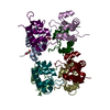
PDBj- Assembly
| Deposited unit | 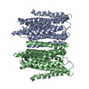
|
|---|---|
| 1 |
|
-Components
| #1: Protein | Mass: 73352.297 Da / Num. of mol.: 2 Source method: isolated from a genetically manipulated source Source: (gene. exp.) Gene: otop3, LOC100127796 / Production host:  Homo sapiens (human) / References: UniProt: A9JTM7 Homo sapiens (human) / References: UniProt: A9JTM7Has protein modification | N | |
|---|
-Experimental details
-Experiment
| Experiment | Method: ELECTRON MICROSCOPY |
|---|---|
| EM experiment | Aggregation state: PARTICLE / 3D reconstruction method: single particle reconstruction |
- Sample preparation
| Component | Name: Xenopus tropicalis OTOP3 / Type: ORGANELLE OR CELLULAR COMPONENT / Entity ID: all / Source: RECOMBINANT |
|---|---|
| Molecular weight | Value: 0.077 MDa / Experimental value: NO |
| Source (natural) | Organism: |
| Source (recombinant) | Organism: Homo sapiens (human) / Cell: HEK293F / Plasmid: pEZT |
| Buffer solution | pH: 8 Details: Solutions were made fresh from stock solutions and filtered. |
| Buffer component | Conc.: 150 mM / Name: sodium chloride / Formula: NaCl |
| Specimen | Conc.: 1.3 mg/ml / Embedding applied: NO / Shadowing applied: NO / Staining applied: NO / Vitrification applied: YES / Details: This sample was monodisperse |
| Vitrification | Instrument: FEI VITROBOT MARK IV / Cryogen name: ETHANE / Humidity: 100 % / Chamber temperature: 277 K |
- Electron microscopy imaging
| Experimental equipment |  Model: Titan Krios / Image courtesy: FEI Company |
|---|---|
| Microscopy | Model: FEI TITAN KRIOS |
| Electron gun | Electron source:  FIELD EMISSION GUN / Accelerating voltage: 300 kV / Illumination mode: FLOOD BEAM FIELD EMISSION GUN / Accelerating voltage: 300 kV / Illumination mode: FLOOD BEAM |
| Electron lens | Mode: BRIGHT FIELD / Nominal magnification: 46730 X / Nominal defocus max: 3000 nm / Nominal defocus min: 1200 nm / Cs: 2.7 mm / C2 aperture diameter: 70 µm / Alignment procedure: COMA FREE |
| Specimen holder | Cryogen: NITROGEN / Specimen holder model: FEI TITAN KRIOS AUTOGRID HOLDER |
| Image recording | Average exposure time: 15 sec. / Electron dose: 50 e/Å2 / Detector mode: SUPER-RESOLUTION / Film or detector model: GATAN K2 SUMMIT (4k x 4k) / Num. of grids imaged: 1 / Num. of real images: 3000 |
| EM imaging optics | Energyfilter name: GIF Quantum LS |
| Image scans | Movie frames/image: 30 |
- Processing
| Software | Name: PHENIX / Version: 1.14_3260: / Classification: refinement | ||||||||||||||||||||||||||||||||
|---|---|---|---|---|---|---|---|---|---|---|---|---|---|---|---|---|---|---|---|---|---|---|---|---|---|---|---|---|---|---|---|---|---|
| EM software |
| ||||||||||||||||||||||||||||||||
| Image processing | Details: 30 frames per movie stack were saved for motion correction. | ||||||||||||||||||||||||||||||||
| CTF correction | Details: The CTF correction was performed during the map refinement in RELION. Type: PHASE FLIPPING AND AMPLITUDE CORRECTION | ||||||||||||||||||||||||||||||||
| Particle selection | Num. of particles selected: 1433949 / Details: The particles were auto-picked in RELION. | ||||||||||||||||||||||||||||||||
| 3D reconstruction | Resolution: 3.92 Å / Resolution method: FSC 0.143 CUT-OFF / Num. of particles: 212551 / Symmetry type: POINT |
