 Movie
Movie Controller

[English] 日本語
 Yorodumi
- PDB-6nym: Helicobacter pylori Vacuolating Cytotoxin A Oligomeric Assembly 2... -
+ Open data

- Basic information
| Entry | Database: PDB / ID: 6nym | ||||||||||||
|---|---|---|---|---|---|---|---|---|---|---|---|---|---|
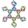
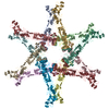
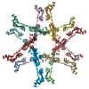
| Title | Helicobacter pylori Vacuolating Cytotoxin A Oligomeric Assembly 2d (OA-2d) | ||||||||||||
 Components Components | Vacuolating cytotoxin autotransporter | ||||||||||||
 Keywords Keywords | TOXIN / Helicobacter pylori / vacuolating cytotoxin A / pore-forming toxin | ||||||||||||
| Function / homology |  Function and homology information Function and homology informationcell outer membrane / toxin activity / periplasmic space / cell surface / extracellular region Similarity search - Function | ||||||||||||
| Biological species |   Helicobacter pylori (bacteria) Helicobacter pylori (bacteria) | ||||||||||||
| Method | ELECTRON MICROSCOPY / single particle reconstruction / cryo EM / Resolution: 3.6 Å | ||||||||||||
 Authors Authors | Zhang, K. / Zhang, H. / Li, S. / Au, S. / Chiu, W. | ||||||||||||
| Funding support |  United States, 3items United States, 3items
| ||||||||||||
 Citation Citation | Journal: Proc Natl Acad Sci U S A / Year: 2019 Title: Cryo-EM structures of vacuolating cytotoxin A oligomeric assemblies at near-atomic resolution. Authors: Kaiming Zhang / Huawei Zhang / Shanshan Li / Grigore D Pintilie / Tung-Chung Mou / Yuanzhu Gao / Qinfen Zhang / Henry van den Bedem / Michael F Schmid / Shannon Wing Ngor Au / Wah Chiu /  Abstract: Human gastric pathogen () is the primary risk factor for gastric cancer and is one of the most prevalent carcinogenic infectious agents. Vacuolating cytotoxin A (VacA) is a key virulence factor ...Human gastric pathogen () is the primary risk factor for gastric cancer and is one of the most prevalent carcinogenic infectious agents. Vacuolating cytotoxin A (VacA) is a key virulence factor secreted by and induces multiple cellular responses. Although structural and functional studies of VacA have been extensively performed, the high-resolution structure of a full-length VacA protomer and the molecular basis of its oligomerization are still unknown. Here, we use cryoelectron microscopy to resolve 10 structures of VacA assemblies, including monolayer (hexamer and heptamer) and bilayer (dodecamer, tridecamer, and tetradecamer) oligomers. The models of the 88-kDa full-length VacA protomer derived from the near-atomic resolution maps are highly conserved among different oligomers and show a continuous right-handed β-helix made up of two domains with extensive domain-domain interactions. The specific interactions between adjacent protomers in the same layer stabilizing the oligomers are well resolved. For double-layer oligomers, we found short- and/or long-range hydrophobic interactions between protomers across the two layers. Our structures and other previous observations lead to a mechanistic model wherein VacA hexamer would correspond to the prepore-forming state, and the N-terminal region of VacA responsible for the membrane insertion would undergo a large conformational change to bring the hydrophobic transmembrane region to the center of the oligomer for the membrane channel formation. | ||||||||||||
| History |
|
- Structure visualization
| Movie |
Movie viewer |
|---|---|
| Structure viewer | Molecule: MolmilJmol/JSmol |
- Downloads & links
-Download
| PDBx/mmCIF format | 6nym.cif.gz | 1.4 MB | Display | PDBx/mmCIF format |
|---|---|---|---|---|
| PDB format | pdb6nym.ent.gz | 1.2 MB | Display | PDB format |
| PDBx/mmJSON format | 6nym.json.gz | Tree view | PDBx/mmJSON format | |
| Others |  Other downloads Other downloads |
-Validation report
| Arichive directory | https://data.pdbj.org/pub/pdb/validation_reports/ny/6nymftp://data.pdbj.org/pub/pdb/validation_reports/ny/6nym | HTTPS FTP |
|---|
-Related structure data
| Related structure data | 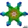 0546MC 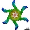 0542C 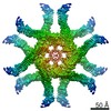 0543C 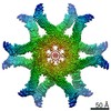 0544C 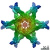 0545C 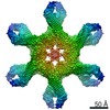 0547C 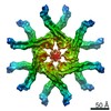 0548C 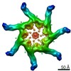 0549C  0550C 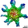 0551C 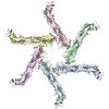 6nyfC 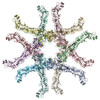 6nygC 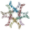 6nyjC 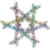 6nylC 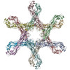 6nynC M: map data used to model this data C: citing same article ( |
|---|---|
| Similar structure data | |
| EM raw data | EMPIAR-10607 (Title: Cryo-EM structures of Helicobacter pylori vacuolating cytotoxin A (VacA) oligomeric assemblies Data size: 3.9 TB Data #1: Helicobacter pylori vacuolating cytotoxin A [micrographs - multiframe]) |
-Links
 PDBj
PDBj- Assembly
| Deposited unit | 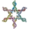
|
|---|---|
| 1 |
|
-Components
| #1: Protein | Mass: 88463.211 Da / Num. of mol.: 12 / Source method: isolated from a natural source / Source: (natural) Helicobacter pylori (bacteria) / References: UniProt: Q48245Has protein modification | Y | |
|---|
-Experimental details
-Experiment
| Experiment | Method: ELECTRON MICROSCOPY |
|---|---|
| EM experiment | Aggregation state: PARTICLE / 3D reconstruction method: single particle reconstruction |
- Sample preparation
| Component | Name: Helicobacter pylori Vacuolating Cytotoxin A Oligomeric Assembly 2d (OA-2d) Type: COMPLEX / Entity ID: all / Source: NATURAL |
|---|---|
| Molecular weight | Value: 0.088 MDa / Experimental value: YES |
| Source (natural) | Organism: Helicobacter pylori (bacteria) / Strain: 60190 |
| Buffer solution | pH: 7.4 |
| Specimen | Conc.: 0.2 mg/ml / Embedding applied: NO / Shadowing applied: NO / Staining applied: NO / Vitrification applied: YES / Details: VacA OA-2d |
| Specimen support | Details: unspecified |
| Vitrification | Instrument: FEI VITROBOT MARK IV / Cryogen name: ETHANE / Humidity: 100 % |
- Electron microscopy imaging
| Experimental equipment |  Model: Titan Krios / Image courtesy: FEI Company |
|---|---|
| Microscopy | Model: FEI TITAN KRIOS |
| Electron gun | Electron source:  FIELD EMISSION GUN / Accelerating voltage: 300 kV / Illumination mode: FLOOD BEAM FIELD EMISSION GUN / Accelerating voltage: 300 kV / Illumination mode: FLOOD BEAM |
| Electron lens | Mode: BRIGHT FIELD / Nominal magnification: 130000 X / Nominal defocus max: 2500 nm / Nominal defocus min: 1300 nm / Cs: 2.7 mm / C2 aperture diameter: 70 µm / Alignment procedure: COMA FREE |
| Specimen holder | Cryogen: NITROGEN / Specimen holder model: FEI TITAN KRIOS AUTOGRID HOLDER |
| Image recording | Average exposure time: 6 sec. / Electron dose: 7 e/Å2 / Detector mode: COUNTING / Film or detector model: GATAN K2 SUMMIT (4k x 4k) / Num. of grids imaged: 3 / Num. of real images: 13708 |
| EM imaging optics | Energyfilter name: GIF Quantum LS / Energyfilter slit width: 20 eV |
| Image scans | Movie frames/image: 30 / Used frames/image: 1-30 |
- Processing
| Software | Name: PHENIX / Version: 1.13_2998: / Classification: refinement | ||||||||||||||||||||||||||||||||||||||||
|---|---|---|---|---|---|---|---|---|---|---|---|---|---|---|---|---|---|---|---|---|---|---|---|---|---|---|---|---|---|---|---|---|---|---|---|---|---|---|---|---|---|
| EM software |
| ||||||||||||||||||||||||||||||||||||||||
| CTF correction | Type: PHASE FLIPPING AND AMPLITUDE CORRECTION | ||||||||||||||||||||||||||||||||||||||||
| Particle selection | Num. of particles selected: 540214 | ||||||||||||||||||||||||||||||||||||||||
| Symmetry | Point symmetry: D6 (2x6 fold dihedral) | ||||||||||||||||||||||||||||||||||||||||
| 3D reconstruction | Resolution: 3.6 Å / Resolution method: FSC 0.143 CUT-OFF / Num. of particles: 31625 / Symmetry type: POINT | ||||||||||||||||||||||||||||||||||||||||
| Atomic model building | Protocol: BACKBONE TRACE | ||||||||||||||||||||||||||||||||||||||||
| Refine LS restraints |
|
