Type: DECTRIS PILATUS3 X 6M / Detector: PIXEL / Date: Dec 9, 2018 / Details: mirrors
Radiation
Monochromator: Si(111) / Protocol: SINGLE WAVELENGTH / Monochromatic (M) / Laue (L): M / Scattering type: x-ray
Radiation wavelength
Wavelength: 0.97926 Å / Relative weight: 1
Reflection
Resolution: 1.4→30 Å / Num. obs: 118279 / % possible obs: 100 % / Observed criterion σ(I): -3 / Redundancy: 9.6 % / Rmerge(I) obs: 0.099 / Net I/σ(I): 24.627
Reflection shell
Resolution: 1.4→1.42 Å / Redundancy: 7.1 % / Rmerge(I) obs: 1.676 / Mean I/σ(I) obs: 1.332 / Num. unique obs: 3040 / CC1/2: 0.709 / % possible all: 99.9
-
Processing
Software
Name
Version
Classification
PHENIX
(dev_2947: ???)
refinement
HKL-3000
datareduction
HKL-3000
datascaling
HKL-3000
phasing
Refinement
Method to determine structure: SAD / Resolution: 1.399→29.197 Å / SU ML: 0.11 / Cross valid method: THROUGHOUT / σ(F): 0.23 / Phase error: 14.88 / Stereochemistry target values: ML
Rfactor
Num. reflection
% reflection
Rfree
0.1512
5758
4.87 %
Rwork
0.1235
-
-
obs
0.1249
118223
99.91 %
Solvent computation
Shrinkage radii: 0.9 Å / VDW probe radii: 1.11 Å / Solvent model: FLAT BULK SOLVENT MODEL
Refinement step
Cycle: LAST / Resolution: 1.399→29.197 Å
Protein
Nucleic acid
Ligand
Solvent
Total
Num. atoms
2011
0
5
215
2231
Refine LS restraints
Refine-ID
Type
Dev ideal
Number
X-RAY DIFFRACTION
f_bond_d
0.012
2085
X-RAY DIFFRACTION
f_angle_d
1.22
2832
X-RAY DIFFRACTION
f_dihedral_angle_d
16.091
776
X-RAY DIFFRACTION
f_chiral_restr
0.092
324
X-RAY DIFFRACTION
f_plane_restr
0.008
369
LS refinement shell
Resolution (Å)
Rfactor Rfree
Num. reflection Rfree
Rfactor Rwork
Num. reflection Rwork
Refine-ID
% reflection obs (%)
1.399-1.4149
0.2622
186
0.2333
3722
X-RAY DIFFRACTION
98
1.4149-1.4315
0.2505
197
0.2103
3700
X-RAY DIFFRACTION
100
1.4315-1.449
0.2279
177
0.21
3780
X-RAY DIFFRACTION
100
1.449-1.4673
0.2546
171
0.1916
3719
X-RAY DIFFRACTION
100
1.4673-1.4866
0.2227
175
0.1885
3868
X-RAY DIFFRACTION
100
1.4866-1.507
0.2167
175
0.173
3707
X-RAY DIFFRACTION
100
1.507-1.5285
0.1931
215
0.1573
3760
X-RAY DIFFRACTION
100
1.5285-1.5513
0.1964
170
0.1465
3753
X-RAY DIFFRACTION
100
1.5513-1.5756
0.1993
209
0.1482
3734
X-RAY DIFFRACTION
100
1.5756-1.6014
0.1714
208
0.1324
3751
X-RAY DIFFRACTION
100
1.6014-1.629
0.1756
185
0.1244
3766
X-RAY DIFFRACTION
100
1.629-1.6586
0.158
180
0.1167
3709
X-RAY DIFFRACTION
100
1.6586-1.6905
0.1575
198
0.11
3766
X-RAY DIFFRACTION
100
1.6905-1.725
0.1348
174
0.1049
3801
X-RAY DIFFRACTION
100
1.725-1.7625
0.1537
218
0.1081
3736
X-RAY DIFFRACTION
100
1.7625-1.8035
0.1567
174
0.1032
3746
X-RAY DIFFRACTION
100
1.8035-1.8486
0.1384
190
0.105
3746
X-RAY DIFFRACTION
100
1.8486-1.8986
0.1506
158
0.0976
3809
X-RAY DIFFRACTION
100
1.8986-1.9545
0.1269
223
0.0969
3689
X-RAY DIFFRACTION
100
1.9545-2.0175
0.118
184
0.099
3790
X-RAY DIFFRACTION
100
2.0175-2.0896
0.1315
206
0.0998
3731
X-RAY DIFFRACTION
100
2.0896-2.1733
0.1327
189
0.1059
3753
X-RAY DIFFRACTION
100
2.1733-2.2721
0.1405
198
0.1076
3739
X-RAY DIFFRACTION
100
2.2721-2.3919
0.1493
242
0.1112
3712
X-RAY DIFFRACTION
100
2.3919-2.5417
0.1379
238
0.1112
3710
X-RAY DIFFRACTION
100
2.5417-2.7378
0.1373
156
0.1208
3772
X-RAY DIFFRACTION
100
2.7378-3.013
0.1567
166
0.1298
3763
X-RAY DIFFRACTION
100
3.013-3.4484
0.1552
210
0.1345
3754
X-RAY DIFFRACTION
100
3.4484-4.3424
0.1374
188
0.113
3720
X-RAY DIFFRACTION
100
4.3424-29.2036
0.1623
198
0.1458
3759
X-RAY DIFFRACTION
100
+
About Yorodumi
-
News
-
Feb 9, 2022. New format data for meta-information of EMDB entries
New format data for meta-information of EMDB entries
Version 3 of the EMDB header file is now the official format.
The previous official version 1.9 will be removed from the archive.
In the structure databanks used in Yorodumi, some data are registered as the other names, "COVID-19 virus" and "2019-nCoV". Here are the details of the virus and the list of structure data.
Jan 31, 2019. EMDB accession codes are about to change! (news from PDBe EMDB page)
EMDB accession codes are about to change! (news from PDBe EMDB page)
The allocation of 4 digits for EMDB accession codes will soon come to an end. Whilst these codes will remain in use, new EMDB accession codes will include an additional digit and will expand incrementally as the available range of codes is exhausted. The current 4-digit format prefixed with “EMD-” (i.e. EMD-XXXX) will advance to a 5-digit format (i.e. EMD-XXXXX), and so on. It is currently estimated that the 4-digit codes will be depleted around Spring 2019, at which point the 5-digit format will come into force.
The EM Navigator/Yorodumi systems omit the EMD- prefix.
Related info.:Q: What is EMD? / ID/Accession-code notation in Yorodumi/EM Navigator
Yorodumi is a browser for structure data from EMDB, PDB, SASBDB, etc.
This page is also the successor to EM Navigator detail page, and also detail information page/front-end page for Omokage search.
The word "yorodu" (or yorozu) is an old Japanese word meaning "ten thousand". "mi" (miru) is to see.
Related info.:EMDB / PDB / SASBDB / Comparison of 3 databanks / Yorodumi Search / Aug 31, 2016. New EM Navigator & Yorodumi / Yorodumi Papers / Jmol/JSmol / Function and homology information / Changes in new EM Navigator and Yorodumi
 Movie
Movie Controller

 Yorodumi
 Open data
 Basic information
 Components
 Keywords
 Function and homology information
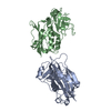
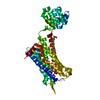
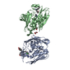
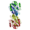
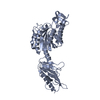
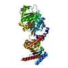
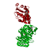
 Clostridium kluyveri (bacteria)
 X-RAY DIFFRACTION /
  Authors
 United States, 1items
  Citation
 Structure visualization
 Downloads & links
 Other downloads

 PDBj
PDBj


 Assembly
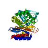

 Mass: 35.453 Da / Num. of mol.: 5 / Source method: obtained synthetically / Formula: Cl
 Mass: 18.015 Da / Num. of mol.: 215 / Source method: isolated from a natural source / Formula: H2O
 Sample preparation
 Processing