 Movie
Movie Controller

[English] 日本語
 Yorodumi
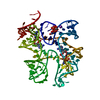
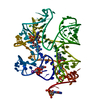
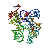
- PDB-6n5o: Structure of Human pir-miRNA-202 Apical Loop and One-base-pair Fu... -
+ Open data

- Basic information
| Entry | Database: PDB / ID: 6n5o | ||||||
|---|---|---|---|---|---|---|---|
| Title | Structure of Human pir-miRNA-202 Apical Loop and One-base-pair Fused to the YdaO Riboswitch Scaffold | ||||||
 Components Components | RNA (126-MER) | ||||||
 Keywords Keywords | RNA / microRNA / RNA processing / Protein-RNA interaction | ||||||
| Function / homology | Chem-2BA / : / RNA / RNA (> 10) / RNA (> 100) Function and homology information Function and homology information | ||||||
| Biological species |  Homo sapiens (human) Homo sapiens (human) | ||||||
| Method |  X-RAY DIFFRACTION / SYNCHROTRON / MOLECULAR REPLACEMENT / Resolution: 2.708 Å X-RAY DIFFRACTION / SYNCHROTRON / MOLECULAR REPLACEMENT / Resolution: 2.708 Å | ||||||
 Authors Authors | Shoffner, G.M. / Peng, Z. / Guo, F. | ||||||
| Funding support |  United States, 1items United States, 1items
| ||||||
 Citation Citation | Journal: To Be Published Title: Three-dimensional structures of pri-miRNA apical junctions and loops revealed by scaffold-directed crystallography Authors: Shoffner, G.M. / Peng, Z. / Guo, F. | ||||||
| History |
|
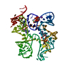
- Structure visualization
| Structure viewer | Molecule: MolmilJmol/JSmol |
|---|
- Downloads & links
-Download
| PDBx/mmCIF format | 6n5o.cif.gz | 153.3 KB | Display | PDBx/mmCIF format |
|---|---|---|---|---|
| PDB format | pdb6n5o.ent.gz | 120.5 KB | Display | PDB format |
| PDBx/mmJSON format | 6n5o.json.gz | Tree view | PDBx/mmJSON format | |
| Others |  Other downloads Other downloads |
-Validation report
| Arichive directory | https://data.pdbj.org/pub/pdb/validation_reports/n5/6n5oftp://data.pdbj.org/pub/pdb/validation_reports/n5/6n5o | HTTPS FTP |
|---|
-Related structure data
| Related structure data |  6n5kC  6n5nC  6n5pC  6n5qC  6n5sC  6n5tC  6wtlC  4qk8S C: citing same article ( S: Starting model for refinement |
|---|---|
| Similar structure data |

-Links
 PDBj
PDBj


- Assembly
| Deposited unit | 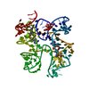
| ||||||||
|---|---|---|---|---|---|---|---|---|---|
| 1 |
| ||||||||
| Unit cell |
|
-Components
-RNA chain , 1 types, 1 molecules A
| #1: RNA chain | Mass: 40915.195 Da / Num. of mol.: 1 Source method: isolated from a genetically manipulated source Source: (gene. exp.) Homo sapiens (human)Production host: in vitro transcription vector pT7-Fluc(deltai) (others) |
|---|
-Non-polymers , 5 types, 12 molecules 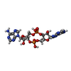


| #2: Chemical |  Mass: 658.412 Da / Num. of mol.: 2 / Source method: obtained synthetically / Formula: C20H24N10O12P2 Mass: 658.412 Da / Num. of mol.: 2 / Source method: obtained synthetically / Formula: C20H24N10O12P2#3: Chemical | ChemComp-MG /  Mass: 24.305 Da / Num. of mol.: 4 / Source method: obtained synthetically / Formula: Mg Mass: 24.305 Da / Num. of mol.: 4 / Source method: obtained synthetically / Formula: Mg#4: Chemical | ChemComp-K / |  Mass: 39.098 Da / Num. of mol.: 1 / Source method: obtained synthetically / Formula: K Mass: 39.098 Da / Num. of mol.: 1 / Source method: obtained synthetically / Formula: K#5: Chemical | ChemComp-SO4 / |  Mass: 96.063 Da / Num. of mol.: 1 / Source method: obtained synthetically / Formula: SO4 Mass: 96.063 Da / Num. of mol.: 1 / Source method: obtained synthetically / Formula: SO4#6: Water | ChemComp-HOH / | Mass: 18.015 Da / Num. of mol.: 4 / Source method: isolated from a natural source / Formula: H2O |
|---|
-Experimental details
-Experiment
| Experiment | Method: X-RAY DIFFRACTION / Number of used crystals: 1 |
|---|
- Sample preparation
| Crystal | Density Matthews: 5.37 Å3/Da / Density % sol: 77.11 % |
|---|---|
| Crystal grow | Temperature: 297 K / Method: vapor diffusion, hanging drop / pH: 7.4 Details: 1.9 M (NH4)2SO4, 0.2 M Li2SO4, and 0.1 M HEPES pH 7.4 Temp details: Room Temperature |
-Data collection
| Diffraction | Mean temperature: 100 K / Serial crystal experiment: N |
|---|---|
| Diffraction source | Source: SYNCHROTRON / Site: APS / Beamline: 24-ID-C / Wavelength: 0.9202 Å |
| Detector | Type: ADSC QUANTUM 315 / Detector: CCD / Date: Oct 25, 2017 |
| Radiation | Monochromator: Cryo-Cooled double crystal / Protocol: SINGLE WAVELENGTH / Monochromatic (M) / Laue (L): M / Scattering type: x-ray |
| Radiation wavelength | Wavelength: 0.9202 Å / Relative weight: 1 |
| Reflection | Resolution: 2.708→99.524 Å / Num. obs: 24343 / % possible obs: 99.9 % / Redundancy: 56.4 % / CC1/2: 0.996 / Rpim(I) all: 0.014 / Rrim(I) all: 0.0106 / Net I/σ(I): 29.9 |
| Reflection shell | Resolution: 2.71→2.81 Å / Redundancy: 39.1 % / Mean I/σ(I) obs: 2.1 / Num. unique obs: 2434 / CC1/2: 0.815 / Rpim(I) all: 0.336 / Rrim(I) all: 2.17 / % possible all: 99 |
- Processing
| Software |
| ||||||||||||||||||||||||||||||||||||||||||||||||||||||||||||||||||||||||||||||||||||||||||||||||||||||||||||||||||||||||||||||
|---|---|---|---|---|---|---|---|---|---|---|---|---|---|---|---|---|---|---|---|---|---|---|---|---|---|---|---|---|---|---|---|---|---|---|---|---|---|---|---|---|---|---|---|---|---|---|---|---|---|---|---|---|---|---|---|---|---|---|---|---|---|---|---|---|---|---|---|---|---|---|---|---|---|---|---|---|---|---|---|---|---|---|---|---|---|---|---|---|---|---|---|---|---|---|---|---|---|---|---|---|---|---|---|---|---|---|---|---|---|---|---|---|---|---|---|---|---|---|---|---|---|---|---|---|---|---|---|
| Refinement | Method to determine structure: MOLECULAR REPLACEMENT Starting model: 4QK8 Resolution: 2.708→99.524 Å / SU ML: 0.39 / Cross valid method: THROUGHOUT / σ(F): 1.37 / Phase error: 24.72
| ||||||||||||||||||||||||||||||||||||||||||||||||||||||||||||||||||||||||||||||||||||||||||||||||||||||||||||||||||||||||||||||
| Solvent computation | Shrinkage radii: 0.9 Å / VDW probe radii: 1.11 Å | ||||||||||||||||||||||||||||||||||||||||||||||||||||||||||||||||||||||||||||||||||||||||||||||||||||||||||||||||||||||||||||||
| Displacement parameters | Biso max: 349.1 Å2 / Biso mean: 113.8796 Å2 / Biso min: 48.19 Å2 | ||||||||||||||||||||||||||||||||||||||||||||||||||||||||||||||||||||||||||||||||||||||||||||||||||||||||||||||||||||||||||||||
| Refinement step | Cycle: final / Resolution: 2.708→99.524 Å
| ||||||||||||||||||||||||||||||||||||||||||||||||||||||||||||||||||||||||||||||||||||||||||||||||||||||||||||||||||||||||||||||
| LS refinement shell | Refine-ID: X-RAY DIFFRACTION / Rfactor Rfree error: 0 / Total num. of bins used: 17
| ||||||||||||||||||||||||||||||||||||||||||||||||||||||||||||||||||||||||||||||||||||||||||||||||||||||||||||||||||||||||||||||
| Refinement TLS params. | Method: refined / Origin x: -76.678 Å / Origin y: 12.7885 Å / Origin z: -17.5249 Å
| ||||||||||||||||||||||||||||||||||||||||||||||||||||||||||||||||||||||||||||||||||||||||||||||||||||||||||||||||||||||||||||||
| Refinement TLS group | Selection details: resid 14 |
