 Movie
Movie Controller

[English] 日本語
 Yorodumi
- PDB-6mwc: CryoEM structure of chimeric Eastern Equine Encephalitis Virus wi... -
+ Open data

- Basic information
| Entry | Database: PDB / ID: 6mwc | |||||||||||||||
|---|---|---|---|---|---|---|---|---|---|---|---|---|---|---|---|---|
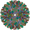
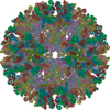
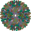
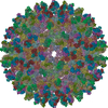
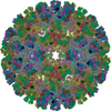
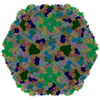
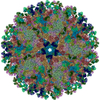
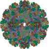
| Title | CryoEM structure of chimeric Eastern Equine Encephalitis Virus with Fab of EEEV-5 antibody | |||||||||||||||
 Components Components |
| |||||||||||||||
 Keywords Keywords | VIRUS/IMMUNE SYSTEM / Alphavirus / EEEV / Eastern Equine Encephalitis Virus / Sindbis / Fab / VIRUS-IMMUNE SYSTEM complex | |||||||||||||||
| Function / homology |  Function and homology information Function and homology informationtogavirin / T=4 icosahedral viral capsid / immunoglobulin complex / symbiont-mediated suppression of host toll-like receptor signaling pathway / host cell cytoplasm / adaptive immune response / symbiont-mediated suppression of host gene expression / serine-type endopeptidase activity / fusion of virus membrane with host endosome membrane / symbiont entry into host cell ...togavirin / T=4 icosahedral viral capsid / immunoglobulin complex / symbiont-mediated suppression of host toll-like receptor signaling pathway / host cell cytoplasm / adaptive immune response / symbiont-mediated suppression of host gene expression / serine-type endopeptidase activity / fusion of virus membrane with host endosome membrane / symbiont entry into host cell / virion attachment to host cell / host cell nucleus / host cell plasma membrane / virion membrane / structural molecule activity / proteolysis / RNA binding Similarity search - Function | |||||||||||||||
| Biological species |  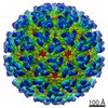 Eastern equine encephalitis virus Eastern equine encephalitis virus | |||||||||||||||
| Method | ELECTRON MICROSCOPY / single particle reconstruction / cryo EM / Resolution: 7.5 Å | |||||||||||||||
 Authors Authors | Hasan, S.S. / Sun, C. / Kim, A.S. / Watanabe, Y. / Chen, C.L. / Klose, T. / Buda, G. / Crispin, M. / Diamond, M.S. / Klimstra, W.B. / Rossmann, M.G. | |||||||||||||||
| Funding support |  United States, United States,  United Kingdom, 4items United Kingdom, 4items
| |||||||||||||||
 Citation Citation | Journal: Cell Rep / Year: 2018 Title: Cryo-EM Structures of Eastern Equine Encephalitis Virus Reveal Mechanisms of Virus Disassembly and Antibody Neutralization. Authors: S Saif Hasan / Chengqun Sun / Arthur S Kim / Yasunori Watanabe / Chun-Liang Chen / Thomas Klose / Geeta Buda / Max Crispin / Michael S Diamond / William B Klimstra / Michael G Rossmann / Abstract: Alphaviruses are enveloped pathogens that cause arthritis and encephalitis. Here, we report a 4.4-Å cryoelectron microscopy (cryo-EM) structure of eastern equine encephalitis virus (EEEV), an ...Alphaviruses are enveloped pathogens that cause arthritis and encephalitis. Here, we report a 4.4-Å cryoelectron microscopy (cryo-EM) structure of eastern equine encephalitis virus (EEEV), an alphavirus that causes fatal encephalitis in humans. Our analysis provides insights into viral entry into host cells. The envelope protein E2 showed a binding site for the cellular attachment factor heparan sulfate. The presence of a cryptic E2 glycan suggests how EEEV escapes surveillance by lectin-expressing myeloid lineage cells, which are sentinels of the immune system. A mechanism for nucleocapsid core release and disassembly upon viral entry was inferred based on pH changes and capsid dissociation from envelope proteins. The EEEV capsid structure showed a viral RNA genome binding site adjacent to a ribosome binding site for viral genome translation following genome release. Using five Fab-EEEV complexes derived from neutralizing antibodies, our investigation provides insights into EEEV host cell interactions and protective epitopes relevant to vaccine design. | |||||||||||||||
| History |
|
- Structure visualization
| Movie |
Movie viewer |
|---|---|
| Structure viewer | Molecule: MolmilJmol/JSmol |
- Downloads & links
-Download
| PDBx/mmCIF format | 6mwc.cif.gz | 884.5 KB | Display | PDBx/mmCIF format |
|---|---|---|---|---|
| PDB format | pdb6mwc.ent.gz | 707.7 KB | Display | PDB format |
| PDBx/mmJSON format | 6mwc.json.gz | Tree view | PDBx/mmJSON format | |
| Others |  Other downloads Other downloads |
-Validation report
| Arichive directory | https://data.pdbj.org/pub/pdb/validation_reports/mw/6mwcftp://data.pdbj.org/pub/pdb/validation_reports/mw/6mwc | HTTPS FTP |
|---|
-Related structure data
| Related structure data | 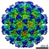 9275MC 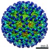 9249C  9274C 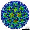 9278C 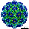 9279C 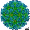 9280C  9281C 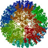 6muiC 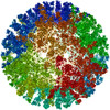 6mw9C  6mwvC 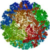 6mwxC  6mx4C 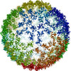 6mx7C M: map data used to model this data C: citing same article ( |
|---|---|
| Similar structure data |
-Links
 PDBj
PDBj


- Assembly
| Deposited unit | 
|
|---|---|
| 1 | x 60
|
| 2 |
|
| 3 | x 5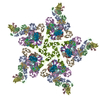
|
| 4 | x 6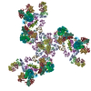
|
| 5 | 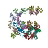
|
| Symmetry | Point symmetry: (Schoenflies symbol: I (icosahedral)) |
-Components
| #1: Protein | Mass: 47938.141 Da / Num. of mol.: 4 / Fragment: ectodomain (UNP residues 802-1242) Source method: isolated from a genetically manipulated source Source: (gene. exp.) Eastern equine encephalitis virus / Cell (production host): BHK-15 / Production host: Mesocricetus auratus (golden hamster) / References: UniProt: E9KXM2, UniProt: Q4QXJ7*PLUS#2: Protein | Mass: 47046.953 Da / Num. of mol.: 4 / Fragment: ectodomain (UNP residues 325-744) Source method: isolated from a genetically manipulated source Source: (gene. exp.) Eastern equine encephalitis virus / Cell (production host): BHK-15 / Production host: Mesocricetus auratus (golden hamster) / References: UniProt: E9KXL2, UniProt: Q4QXJ7*PLUS#3: Antibody | Mass: 23743.719 Da / Num. of mol.: 4 / Fragment: Fab / Source method: isolated from a natural source / Source: (natural) #4: Antibody | Mass: 23177.713 Da / Num. of mol.: 4 / Fragment: Fab / Source method: isolated from a natural source / Source: (natural) Has protein modification | Y | Sequence details | The antibody heavy chain and light chain amino acid sequences in this model are Fab CHK265 (from ...The antibody heavy chain and light chain amino acid sequences in this model are Fab CHK265 (from PDB entry 5ANY) and do not correspond to the amino acid sequences of the antibody heavy chain and light chains present in the sample. | |
|---|
-Experimental details
-Experiment
| Experiment | Method: ELECTRON MICROSCOPY |
|---|---|
| EM experiment | Aggregation state: PARTICLE / 3D reconstruction method: single particle reconstruction |
- Sample preparation
| Component |
| ||||||||||||||||||||||||||||
|---|---|---|---|---|---|---|---|---|---|---|---|---|---|---|---|---|---|---|---|---|---|---|---|---|---|---|---|---|---|
| Source (natural) |
| ||||||||||||||||||||||||||||
| Source (recombinant) | Organism: Mesocricetus auratus (golden hamster) / Cell: BHK-15 | ||||||||||||||||||||||||||||
| Details of virus | Empty: NO / Enveloped: YES / Isolate: OTHER / Type: VIRION | ||||||||||||||||||||||||||||
| Buffer solution | pH: 7.5 | ||||||||||||||||||||||||||||
| Specimen | Conc.: 1 mg/ml / Embedding applied: NO / Shadowing applied: NO / Staining applied: NO / Vitrification applied: YES | ||||||||||||||||||||||||||||
| Specimen support | Details: unspecified | ||||||||||||||||||||||||||||
| Vitrification | Instrument: GATAN CRYOPLUNGE 3 / Cryogen name: HELIUM / Humidity: 80 % / Chamber temperature: 295 K |
- Electron microscopy imaging
| Experimental equipment |  Model: Titan Krios / Image courtesy: FEI Company |
|---|---|
| Microscopy | Model: FEI TITAN KRIOS |
| Electron gun | Electron source:  FIELD EMISSION GUN / Accelerating voltage: 300 kV / Illumination mode: FLOOD BEAM FIELD EMISSION GUN / Accelerating voltage: 300 kV / Illumination mode: FLOOD BEAM |
| Electron lens | Mode: BRIGHT FIELD |
| Image recording | Electron dose: 31 e/Å2 / Film or detector model: GATAN K2 SUMMIT (4k x 4k) |
- Processing
| CTF correction | Type: PHASE FLIPPING AND AMPLITUDE CORRECTION |
|---|---|
| 3D reconstruction | Resolution: 7.5 Å / Resolution method: FSC 0.143 CUT-OFF / Num. of particles: 6583 / Symmetry type: POINT |
| Atomic model building | Protocol: RIGID BODY FIT |
