 Movie
Movie Controller

[English] 日本語
 Yorodumi
- PDB-6mvy: NavAb voltage-gated sodium channel, residues 1-226, crystallized ... -
+ Open data

- Basic information
| Entry | Database: PDB / ID: 6mvy | ||||||
|---|---|---|---|---|---|---|---|
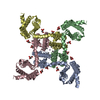
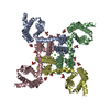
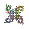
| Title | NavAb voltage-gated sodium channel, residues 1-226, crystallized in the presence of Class 1B Anti-arrhythmic drug Lidocaine | ||||||
 Components Components | Ion transport protein | ||||||
 Keywords Keywords | MEMBRANE PROTEIN / Ion channel Voltage-gated Sodium Channel | ||||||
| Function / homology |  Function and homology information Function and homology informationvoltage-gated sodium channel complex / voltage-gated sodium channel activity / metal ion binding / identical protein binding Similarity search - Function | ||||||
| Biological species |  Arcobacter butzleri (bacteria) Arcobacter butzleri (bacteria) | ||||||
| Method |  X-RAY DIFFRACTION / SYNCHROTRON / MOLECULAR REPLACEMENT / Resolution: 3.002 Å X-RAY DIFFRACTION / SYNCHROTRON / MOLECULAR REPLACEMENT / Resolution: 3.002 Å | ||||||
 Authors Authors | Lenaeus, M.J. / Catterall, W.A. | ||||||
 Citation Citation | Journal: Proc. Natl. Acad. Sci. U.S.A. / Year: 2018 Title: Fenestrations control resting-state block of a voltage-gated sodium channel. Authors: Gamal El-Din, T.M. / Lenaeus, M.J. / Zheng, N. / Catterall, W.A. | ||||||
| History |
|
- Structure visualization
| Structure viewer | Molecule: MolmilJmol/JSmol |
|---|
- Downloads & links
-Download
| PDBx/mmCIF format | 6mvy.cif.gz | 203.4 KB | Display | PDBx/mmCIF format |
|---|---|---|---|---|
| PDB format | pdb6mvy.ent.gz | 164.3 KB | Display | PDB format |
| PDBx/mmJSON format | 6mvy.json.gz | Tree view | PDBx/mmJSON format | |
| Others |  Other downloads Other downloads |
-Validation report
| Arichive directory | https://data.pdbj.org/pub/pdb/validation_reports/mv/6mvyftp://data.pdbj.org/pub/pdb/validation_reports/mv/6mvy | HTTPS FTP |
|---|
-Related structure data
| Related structure data |  6mvvC  6mvwC  6mvxC  3rvyS C: citing same article ( S: Starting model for refinement |
|---|---|
| Similar structure data |
-Links
 PDBj
PDBj

- Assembly
| Deposited unit | 
| ||||||||
|---|---|---|---|---|---|---|---|---|---|
| 1 | 
| ||||||||
| 2 | 
| ||||||||
| Unit cell |
|
-Components
| #1: Protein | Mass: 28202.602 Da / Num. of mol.: 2 / Mutation: I217C Source method: isolated from a genetically manipulated source Source: (gene. exp.) Arcobacter butzleri (strain RM4018) (bacteria)Strain: RM4018 / Gene: Abu_1752 / Production host:  Trichoplusia ni (cabbage looper) / References: UniProt: A8EVM5 Trichoplusia ni (cabbage looper) / References: UniProt: A8EVM5#2: Chemical | ChemComp-PX4 /   Mass: 678.940 Da / Num. of mol.: 8 / Source method: obtained synthetically / Formula: C36H73NO8P / Comment: DMPC, phospholipid*YM Mass: 678.940 Da / Num. of mol.: 8 / Source method: obtained synthetically / Formula: C36H73NO8P / Comment: DMPC, phospholipid*YM#3: Chemical |   Mass: 96.063 Da / Num. of mol.: 2 / Source method: obtained synthetically / Formula: SO4 Mass: 96.063 Da / Num. of mol.: 2 / Source method: obtained synthetically / Formula: SO4#4: Water | ChemComp-HOH / |  Mass: 18.015 Da / Num. of mol.: 39 / Source method: isolated from a natural source / Formula: H2O Mass: 18.015 Da / Num. of mol.: 39 / Source method: isolated from a natural source / Formula: H2O |
|---|
-Experimental details
-Experiment
| Experiment | Method: X-RAY DIFFRACTION / Number of used crystals: 1 |
|---|
- Sample preparation
| Crystal | Density Matthews: 6.83 Å3/Da / Density % sol: 81.98 % |
|---|---|
| Crystal grow | Temperature: 277 K / Method: vapor diffusion, hanging drop Details: 1.8 M Ammonium Sulfate 100 mM Sodium Acetate, pH 5.0 10 mM Lidocaine |
-Data collection
| Diffraction | Mean temperature: 100 K / Serial crystal experiment: N |
|---|---|
| Diffraction source | Source: SYNCHROTRON / Site: ALS  / Beamline: 8.2.1 / Wavelength: 1 Å / Beamline: 8.2.1 / Wavelength: 1 Å |
| Detector | Type: ADSC QUANTUM 315 / Detector: CCD / Date: May 8, 2015 |
| Radiation | Protocol: SINGLE WAVELENGTH / Monochromatic (M) / Laue (L): M / Scattering type: x-ray |
| Radiation wavelength | Wavelength: 1 Å / Relative weight: 1 |
| Reflection | Resolution: 3→50 Å / Num. obs: 30052 / % possible obs: 100 % / Redundancy: 16 % / Rmerge(I) obs: 0.12 / Net I/σ(I): 24 |
| Reflection shell | Resolution: 3.002→3.109 Å / Num. unique all: 2887 / CC1/2: 0.657 |
- Processing
| Software |
| ||||||||||||||||||||||||||||||||||||||||||||||||||||||||||||||||||||||||||||||||||||||||||||||||||||||||||||||||||||||||||||||||||||||||||||||||||||||||||||||||||||||||||||||||||||||||||||||||||||||||
|---|---|---|---|---|---|---|---|---|---|---|---|---|---|---|---|---|---|---|---|---|---|---|---|---|---|---|---|---|---|---|---|---|---|---|---|---|---|---|---|---|---|---|---|---|---|---|---|---|---|---|---|---|---|---|---|---|---|---|---|---|---|---|---|---|---|---|---|---|---|---|---|---|---|---|---|---|---|---|---|---|---|---|---|---|---|---|---|---|---|---|---|---|---|---|---|---|---|---|---|---|---|---|---|---|---|---|---|---|---|---|---|---|---|---|---|---|---|---|---|---|---|---|---|---|---|---|---|---|---|---|---|---|---|---|---|---|---|---|---|---|---|---|---|---|---|---|---|---|---|---|---|---|---|---|---|---|---|---|---|---|---|---|---|---|---|---|---|---|---|---|---|---|---|---|---|---|---|---|---|---|---|---|---|---|---|---|---|---|---|---|---|---|---|---|---|---|---|---|---|---|---|
| Refinement | Method to determine structure: MOLECULAR REPLACEMENT Starting model: 3RVY Resolution: 3.002→40.033 Å / SU ML: 0.39 / Cross valid method: FREE R-VALUE / σ(F): 1.36 / Phase error: 26.12
| ||||||||||||||||||||||||||||||||||||||||||||||||||||||||||||||||||||||||||||||||||||||||||||||||||||||||||||||||||||||||||||||||||||||||||||||||||||||||||||||||||||||||||||||||||||||||||||||||||||||||
| Solvent computation | Shrinkage radii: 0.9 Å / VDW probe radii: 1.11 Å | ||||||||||||||||||||||||||||||||||||||||||||||||||||||||||||||||||||||||||||||||||||||||||||||||||||||||||||||||||||||||||||||||||||||||||||||||||||||||||||||||||||||||||||||||||||||||||||||||||||||||
| Refinement step | Cycle: LAST / Resolution: 3.002→40.033 Å
| ||||||||||||||||||||||||||||||||||||||||||||||||||||||||||||||||||||||||||||||||||||||||||||||||||||||||||||||||||||||||||||||||||||||||||||||||||||||||||||||||||||||||||||||||||||||||||||||||||||||||
| Refine LS restraints |
| ||||||||||||||||||||||||||||||||||||||||||||||||||||||||||||||||||||||||||||||||||||||||||||||||||||||||||||||||||||||||||||||||||||||||||||||||||||||||||||||||||||||||||||||||||||||||||||||||||||||||
| LS refinement shell |
| ||||||||||||||||||||||||||||||||||||||||||||||||||||||||||||||||||||||||||||||||||||||||||||||||||||||||||||||||||||||||||||||||||||||||||||||||||||||||||||||||||||||||||||||||||||||||||||||||||||||||
| Refinement TLS params. | Method: refined / Refine-ID: X-RAY DIFFRACTION
| ||||||||||||||||||||||||||||||||||||||||||||||||||||||||||||||||||||||||||||||||||||||||||||||||||||||||||||||||||||||||||||||||||||||||||||||||||||||||||||||||||||||||||||||||||||||||||||||||||||||||
| Refinement TLS group |
|
