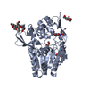
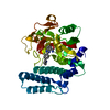
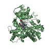
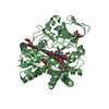
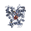
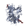
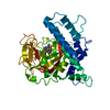
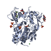
Entry Database : PDB / ID : 6mvdTitle Crystal structure of Lecithin:cholesterol acyltransferase (LCAT) in complex with isopropyl dodec-11-enylfluorophosphonate (IDFP) and a small molecule activator Phosphatidylcholine-sterol acyltransferase Keywords / / / / / / Function / homology Function Domain/homology Component
/ / / / / / / / / / / / / / / / / / / / / / / / / / / / / / / / / / / / / / / / / / / Biological species Homo sapiens (human)Method / / / Resolution : 3.1 Å Authors Manthei, K.A. / Chang, L. / Tesmer, J.J.G. Funding support Organization Grant number Country National Institutes of Health/National Heart, Lung, and Blood Institute (NIH/NHLBI) HL122416 National Institutes of Health/National Heart, Lung, and Blood Institute (NIH/NHLBI) HL131288
Journal : Elife / Year : 2018Title : Molecular basis for activation of lecithin:cholesterol acyltransferase by a compound that increases HDL cholesterol.Authors : Manthei, K.A. / Yang, S.M. / Baljinnyam, B. / Chang, L. / Glukhova, A. / Yuan, W. / Freeman, L.A. / Maloney, D.J. / Schwendeman, A. / Remaley, A.T. / Jadhav, A. / Tesmer, J.J. History Deposition Oct 25, 2018 Deposition site / Processing site Supersession Dec 5, 2018 ID 6DTJ Revision 1.0 Dec 5, 2018 Provider / Type Revision 1.1 Dec 12, 2018 Group / Database references / Category / citation_authorItem _citation.journal_volume / _citation.pdbx_database_id_DOI ... _citation.journal_volume / _citation.pdbx_database_id_DOI / _citation.pdbx_database_id_PubMed / _citation.title / _citation_author.identifier_ORCID / _citation_author.name Revision 1.2 Dec 4, 2019 Group / Category / Item Revision 1.3 Jul 29, 2020 Group / Derived calculations / Structure summaryCategory chem_comp / entity ... chem_comp / entity / pdbx_chem_comp_identifier / pdbx_entity_nonpoly / pdbx_struct_conn_angle / struct_conn / struct_site / struct_site_gen Item _chem_comp.name / _chem_comp.type ... _chem_comp.name / _chem_comp.type / _entity.pdbx_description / _pdbx_entity_nonpoly.name / _pdbx_struct_conn_angle.ptnr1_auth_seq_id / _pdbx_struct_conn_angle.ptnr1_label_atom_id / _pdbx_struct_conn_angle.ptnr1_label_seq_id / _pdbx_struct_conn_angle.ptnr2_symmetry / _pdbx_struct_conn_angle.ptnr3_auth_seq_id / _pdbx_struct_conn_angle.ptnr3_label_atom_id / _pdbx_struct_conn_angle.ptnr3_label_seq_id / _pdbx_struct_conn_angle.value / _struct_conn.conn_type_id / _struct_conn.id / _struct_conn.pdbx_dist_value / _struct_conn.pdbx_leaving_atom_flag / _struct_conn.pdbx_role / _struct_conn.ptnr1_auth_asym_id / _struct_conn.ptnr1_auth_comp_id / _struct_conn.ptnr1_auth_seq_id / _struct_conn.ptnr1_label_asym_id / _struct_conn.ptnr1_label_atom_id / _struct_conn.ptnr1_label_comp_id / _struct_conn.ptnr1_label_seq_id / _struct_conn.ptnr2_auth_asym_id / _struct_conn.ptnr2_auth_comp_id / _struct_conn.ptnr2_auth_seq_id / _struct_conn.ptnr2_label_asym_id / _struct_conn.ptnr2_label_atom_id / _struct_conn.ptnr2_label_comp_id / _struct_conn.ptnr2_symmetry Description / Provider / Type Revision 1.4 Oct 11, 2023 Group Data collection / Database references ... Data collection / Database references / Refinement description / Structure summary Category chem_comp / chem_comp_atom ... chem_comp / chem_comp_atom / chem_comp_bond / database_2 / pdbx_initial_refinement_model Item / _database_2.pdbx_DOI / _database_2.pdbx_database_accessionRevision 1.5 Nov 6, 2024 Group / Category / pdbx_modification_feature
Show all Show less
 Movie
Movie Controller

 Yorodumi
 Open data
 Basic information
 Components
 Keywords
 Function and homology information
 Homo sapiens (human)
 X-RAY DIFFRACTION /
  Authors
 United States, 2items
  Citation
 Structure visualization
 Downloads & links
 Other downloads


 PDBj
PDBj


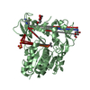
 Assembly
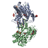

 Type: D-saccharide, beta linking / Mass: 221.208 Da / Num. of mol.: 5


 Mass: 152.129 Da / Num. of mol.: 2 / Source method: obtained synthetically / Formula: C5H13O3P
 Mass: 474.360 Da / Num. of mol.: 2 / Source method: obtained synthetically / Formula: C19H16F6N6O2
 Mass: 96.063 Da / Num. of mol.: 6 / Source method: obtained synthetically / Formula: SO4
 Mass: 58.693 Da / Num. of mol.: 1 / Source method: obtained synthetically / Formula: Ni
 Sample preparation
 Processing