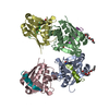
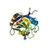
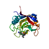
| Deposited unit | A: Double Glycine Motif Protease domain from AMS/PCAT Transporter
B: Double Glycine Motif Protease domain from AMS/PCAT Transporter
C: Double Glycine Motif Protease domain from AMS/PCAT Transporter
D: Double Glycine Motif Protease domain from AMS/PCAT Transporter
M: peptide aldehyde inhibitor 1 based on the ProcA2.8 leader peptide
N: peptide aldehyde inhibitor 1 based on the ProcA2.8 leader peptide
O: peptide aldehyde inhibitor 1 based on the ProcA2.8 leader peptide
P: peptide aldehyde inhibitor 1 based on the ProcA2.8 leader peptide
hetero molecules
| Theoretical mass | Number of molelcules |
|---|
| Total (without water) | 72,865 | 10 |
|---|
| Polymers | 72,276 | 8 |
|---|
| Non-polymers | 589 | 2 |
|---|
| Water | 2,216 | 123 |
|---|
|
|---|
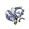
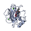
| 1 | A: Double Glycine Motif Protease domain from AMS/PCAT Transporter
M: peptide aldehyde inhibitor 1 based on the ProcA2.8 leader peptide
hetero molecules
| Theoretical mass | Number of molelcules |
|---|
| Total (without water) | 18,363 | 3 |
|---|
| Polymers | 18,069 | 2 |
|---|
| Non-polymers | 294 | 1 |
|---|
| Water | 36 | 2 |
|---|
| Type | Name | Symmetry operation | Number |
|---|
| identity operation | 1_555 | x,y,z | 1 |
| Buried area | 1360 Å2 |
|---|
| ΔGint | -8 kcal/mol |
|---|
| Surface area | 7910 Å2 |
|---|
| Method | PISA |
|---|
|
|---|
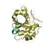
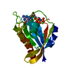
| 2 | B: Double Glycine Motif Protease domain from AMS/PCAT Transporter
N: peptide aldehyde inhibitor 1 based on the ProcA2.8 leader peptide
hetero molecules
| Theoretical mass | Number of molelcules |
|---|
| Total (without water) | 18,363 | 3 |
|---|
| Polymers | 18,069 | 2 |
|---|
| Non-polymers | 294 | 1 |
|---|
| Water | 36 | 2 |
|---|
| Type | Name | Symmetry operation | Number |
|---|
| identity operation | 1_555 | x,y,z | 1 |
| Buried area | 1370 Å2 |
|---|
| ΔGint | -8 kcal/mol |
|---|
| Surface area | 7680 Å2 |
|---|
| Method | PISA |
|---|
|
|---|
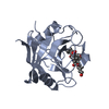
| 3 | C: Double Glycine Motif Protease domain from AMS/PCAT Transporter
O: peptide aldehyde inhibitor 1 based on the ProcA2.8 leader peptide
| Theoretical mass | Number of molelcules |
|---|
| Total (without water) | 18,069 | 2 |
|---|
| Polymers | 18,069 | 2 |
|---|
| Non-polymers | 0 | 0 |
|---|
| Water | 36 | 2 |
|---|
| Type | Name | Symmetry operation | Number |
|---|
| identity operation | 1_555 | x,y,z | 1 |
| Buried area | 1990 Å2 |
|---|
| ΔGint | -5 kcal/mol |
|---|
| Surface area | 7600 Å2 |
|---|
| Method | PISA |
|---|
|
|---|
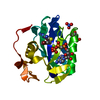
| 4 | D: Double Glycine Motif Protease domain from AMS/PCAT Transporter
P: peptide aldehyde inhibitor 1 based on the ProcA2.8 leader peptide
| Theoretical mass | Number of molelcules |
|---|
| Total (without water) | 18,069 | 2 |
|---|
| Polymers | 18,069 | 2 |
|---|
| Non-polymers | 0 | 0 |
|---|
| Water | 36 | 2 |
|---|
| Type | Name | Symmetry operation | Number |
|---|
| identity operation | 1_555 | x,y,z | 1 |
| Buried area | 1990 Å2 |
|---|
| ΔGint | -5 kcal/mol |
|---|
| Surface area | 7770 Å2 |
|---|
| Method | PISA |
|---|
|
|---|
| Unit cell | | Length a, b, c (Å) | 37.888, 119.426, 76.522 |
|---|
| Angle α, β, γ (deg.) | 90.00, 93.84, 90.00 |
|---|
| Int Tables number | 4 |
|---|
| Space group name H-M | P1211 |
|---|
|
|---|
| Noncrystallographic symmetry (NCS) | NCS domain: | ID | Ens-ID | Details (eV) |
|---|
| 1 | 1 | A| 2 | 1 | B| 1 | 2 | A| 2 | 2 | C| 1 | 3 | A| 2 | 3 | D| 1 | 4 | B| 2 | 4 | C| 1 | 5 | B| 2 | 5 | D| 1 | 6 | C| 2 | 6 | D| 1 | 7 | M| 2 | 7 | N| 1 | 8 | M| 2 | 8 | O| 1 | 9 | M| 2 | 9 | P| 1 | 10 | N| 2 | 10 | O| 1 | 11 | N| 2 | 11 | P| 1 | 12 | O| 2 | 12 | P | | | | | | | | | | | | | | | | | | | | | | | |
NCS domain segments: Component-ID: _ / Refine code: _ | Dom-ID | Ens-ID | Beg auth comp-ID | Beg label comp-ID | End auth comp-ID | End label comp-ID | Auth asym-ID | Label asym-ID | Auth seq-ID | Label seq-ID |
|---|
| 1 | 1 | GLNGLNPROPROAA| 5 - 143 | 5 - 143 | | 2 | 1 | GLNGLNPROPROBB| 5 - 143 | 5 - 143 | | 1 | 2 | GLNGLNPROPROAA| 5 - 143 | 5 - 143 | | 2 | 2 | GLNGLNPROPROCC| 5 - 143 | 5 - 143 | | 1 | 3 | GLNGLNTHRTHRAA| 5 - 144 | 5 - 144 | | 2 | 3 | GLNGLNTHRTHRDD| 5 - 144 | 5 - 144 | | 1 | 4 | GLNGLNTHRTHRBB| 5 - 144 | 5 - 144 | | 2 | 4 | GLNGLNTHRTHRCC| 5 - 144 | 5 - 144 | | 1 | 5 | GLNGLNPROPROBB| 5 - 143 | 5 - 143 | | 2 | 5 | GLNGLNPROPRODD| 5 - 143 | 5 - 143 | | 1 | 6 | GLNGLNPROPROCC| 5 - 143 | 5 - 143 | | 2 | 6 | GLNGLNPROPRODD| 5 - 143 | 5 - 143 | | 1 | 7 | GLYGLYGLYGLYM| E | | | | | | | | | | | | | | | | | | | | | | | | | | | | | | | | | | | | | | | | | | | | | | | | | | | | | | | | | | | | | | | | | | | | | | | | | | | | | |
|
|---|
 Movie
Movie Controller

 Yorodumi
 Open data
 Basic information
 Components
 Keywords
 Function and homology information
 Lachnospiraceae bacterium C6A11 (bacteria)
 X-RAY DIFFRACTION /
  Authors
 United States, 1items
  Citation
 Structure visualization
 Downloads & links
 Other downloads
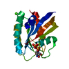


 PDBj
PDBj Assembly