 Movie
Movie Controller

[English] 日本語
 Yorodumi
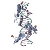
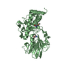
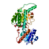
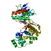
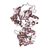
- PDB-6ml2: ZBTB24 Zinc Fingers 4-8 with 19+1mer DNA Oligonucleotide (Sequence 1) -
+ Open data

- Basic information
| Entry | Database: PDB / ID: 6ml2 | ||||||
|---|---|---|---|---|---|---|---|
| Title | ZBTB24 Zinc Fingers 4-8 with 19+1mer DNA Oligonucleotide (Sequence 1) | ||||||
 Components Components |
| ||||||
 Keywords Keywords | TRANSCRIPTION/DNA / protein-DNA complex / transcription / TRANSCRIPTION-DNA complex | ||||||
| Function / homology |  Function and homology information Function and homology informationregulation of immune system process / hematopoietic progenitor cell differentiation / regulation of cytokine production / DNA-binding transcription repressor activity, RNA polymerase II-specific / RNA polymerase II cis-regulatory region sequence-specific DNA binding / DNA-binding transcription factor activity / regulation of DNA-templated transcription / negative regulation of transcription by RNA polymerase II / zinc ion binding / nucleoplasm Similarity search - Function | ||||||
| Biological species |  | ||||||
| Method |  X-RAY DIFFRACTION / SYNCHROTRON / MOLECULAR REPLACEMENT / Resolution: 1.874 Å X-RAY DIFFRACTION / SYNCHROTRON / MOLECULAR REPLACEMENT / Resolution: 1.874 Å | ||||||
 Authors Authors | Horton, J.R. / Cheng, X. / Ren, R. | ||||||
| Funding support |  United States, 1items United States, 1items
| ||||||
 Citation Citation | Journal: Nucleic Acids Res. / Year: 2019 Title: Structural basis of specific DNA binding by the transcription factor ZBTB24. Authors: Ren, R. / Hardikar, S. / Horton, J.R. / Lu, Y. / Zeng, Y. / Singh, A.K. / Lin, K. / Coletta, L.D. / Shen, J. / Lin Kong, C.S. / Hashimoto, H. / Zhang, X. / Chen, T. / Cheng, X. | ||||||
| History |
|
- Structure visualization
| Structure viewer | Molecule: MolmilJmol/JSmol |
|---|
- Downloads & links
-Download
| PDBx/mmCIF format | 6ml2.cif.gz | 67.6 KB | Display | PDBx/mmCIF format |
|---|---|---|---|---|
| PDB format | pdb6ml2.ent.gz | 44.1 KB | Display | PDB format |
| PDBx/mmJSON format | 6ml2.json.gz | Tree view | PDBx/mmJSON format | |
| Others |  Other downloads Other downloads |
-Validation report
| Arichive directory | https://data.pdbj.org/pub/pdb/validation_reports/ml/6ml2ftp://data.pdbj.org/pub/pdb/validation_reports/ml/6ml2 | HTTPS FTP |
|---|
-Related structure data
| Related structure data |  6ml3C  6ml4C  6ml5C  6ml6C  6ml7C  5v3gS S: Starting model for refinement C: citing same article ( |
|---|---|
| Similar structure data |


-Links
 PDBj
PDBj


- Assembly
| Deposited unit | 
| ||||||||
|---|---|---|---|---|---|---|---|---|---|
| 1 |
| ||||||||
| Unit cell |
|
-Components
| #1: Protein | Mass: 17274.033 Da / Num. of mol.: 1 / Fragment: zinc fingers 4-8 (UNP residues 375-519) Source method: isolated from a genetically manipulated source Source: (gene. exp.)  | ||
|---|---|---|---|
| #2: DNA chain | Mass: 6143.981 Da / Num. of mol.: 1 / Source method: obtained synthetically / Source: (synth.) | ||
| #3: DNA chain | Mass: 6125.953 Da / Num. of mol.: 1 / Source method: obtained synthetically / Source: (synth.) | ||
| #4: Chemical | ChemComp-ZN /   Mass: 65.409 Da / Num. of mol.: 5 / Source method: obtained synthetically / Formula: Zn Mass: 65.409 Da / Num. of mol.: 5 / Source method: obtained synthetically / Formula: Zn#5: Water | ChemComp-HOH / |  Mass: 18.015 Da / Num. of mol.: 25 / Source method: isolated from a natural source / Formula: H2O Mass: 18.015 Da / Num. of mol.: 25 / Source method: isolated from a natural source / Formula: H2O |
-Experimental details
-Experiment
| Experiment | Method: X-RAY DIFFRACTION / Number of used crystals: 1 |
|---|
- Sample preparation
| Crystal | Density Matthews: 2.65 Å3/Da / Density % sol: 53.5 % |
|---|---|
| Crystal grow | Temperature: 292 K / Method: vapor diffusion, sitting drop / pH: 6.5 / Details: 25% PEG3350, 0.1 M Bis-Tris, pH 6.5 |
-Data collection
| Diffraction | Mean temperature: 100 K / Serial crystal experiment: N |
|---|---|
| Diffraction source | Source: SYNCHROTRON / Site: APS / Beamline: 22-ID / Wavelength: 1 Å |
| Detector | Type: DECTRIS EIGER X 16M / Detector: PIXEL / Date: Jul 3, 2018 |
| Radiation | Monochromator: Rosenbaum-Rock double crystal Si(111) / Protocol: SINGLE WAVELENGTH / Monochromatic (M) / Laue (L): M / Scattering type: x-ray |
| Radiation wavelength | Wavelength: 1 Å / Relative weight: 1 |
| Reflection | Resolution: 1.87→40.12 Å / Num. obs: 36754 / % possible obs: 77.7 % / Redundancy: 5.6 % / Rmerge(I) obs: 0.056 / Rpim(I) all: 0.024 / Net I/σ(I): 22.6 |
| Reflection shell | Resolution: 1.87→1.94 Å / Redundancy: 3.9 % / Rmerge(I) obs: 0.564 / Mean I/σ(I) obs: 2.1 / Num. unique obs: 699 / CC1/2: 0.864 / Rpim(I) all: 0.264 / % possible all: 27.6 |
- Processing
| Software |
| ||||||||||||||||||||||||||||||||||||||||||||||||||||||||||||||||||||||||||||||||||||||||||||||||||
|---|---|---|---|---|---|---|---|---|---|---|---|---|---|---|---|---|---|---|---|---|---|---|---|---|---|---|---|---|---|---|---|---|---|---|---|---|---|---|---|---|---|---|---|---|---|---|---|---|---|---|---|---|---|---|---|---|---|---|---|---|---|---|---|---|---|---|---|---|---|---|---|---|---|---|---|---|---|---|---|---|---|---|---|---|---|---|---|---|---|---|---|---|---|---|---|---|---|---|---|
| Refinement | Method to determine structure: MOLECULAR REPLACEMENT Starting model: PDB entry 5V3G (zinc fingers) + 18-mer B-DNA model Resolution: 1.874→38.232 Å / SU ML: 0.35 / Cross valid method: THROUGHOUT / σ(F): 1.35 / Phase error: 45.61 / Stereochemistry target values: ML
| ||||||||||||||||||||||||||||||||||||||||||||||||||||||||||||||||||||||||||||||||||||||||||||||||||
| Solvent computation | Shrinkage radii: 0.9 Å / VDW probe radii: 1.11 Å / Solvent model: FLAT BULK SOLVENT MODEL | ||||||||||||||||||||||||||||||||||||||||||||||||||||||||||||||||||||||||||||||||||||||||||||||||||
| Refinement step | Cycle: LAST / Resolution: 1.874→38.232 Å
| ||||||||||||||||||||||||||||||||||||||||||||||||||||||||||||||||||||||||||||||||||||||||||||||||||
| Refine LS restraints |
| ||||||||||||||||||||||||||||||||||||||||||||||||||||||||||||||||||||||||||||||||||||||||||||||||||
| LS refinement shell |
|
