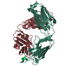
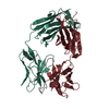
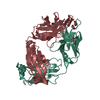
Mass: 23429.818 Da / Num. of mol.: 2 Source method: isolated from a genetically manipulated source Source: (gene. exp.) Homo sapiens (human) / Production host: Escherichia coli (E. coli)
Mass: 18.015 Da / Num. of mol.: 664 / Source method: isolated from a natural source / Formula: H2O
Has protein modification
Y
-
Experimental details
-
Experiment
Experiment
Method: X-RAY DIFFRACTION / Number of used crystals: 1
-
Sample preparation
Crystal
Density Matthews: 2.71 Å3/Da / Density % sol: 54.54 %
Crystal grow
Temperature: 296 K / Method: vapor diffusion, sitting drop Details: Crystals of JTO-FL were grown via sitting-drop vapor diffusion using a crystallization buffer consisting of 20% PEG 3350 and 0.2 M NH4H2PO4 at 23 degrees C. Both diamond-shaped and plate- ...Details: Crystals of JTO-FL were grown via sitting-drop vapor diffusion using a crystallization buffer consisting of 20% PEG 3350 and 0.2 M NH4H2PO4 at 23 degrees C. Both diamond-shaped and plate-shaped crystals were generated in the same drop using these conditions, but only the plate-shaped crystals produced usable diffraction data. Crystals were harvested and immediately flash cooled in liquid nitrogen.
Resolution: 1.75→44.66 Å / Cor.coef. Fo:Fc: 0.973 / Cor.coef. Fo:Fc free: 0.955 / SU B: 3.487 / SU ML: 0.103 / Cross valid method: THROUGHOUT / ESU R: 0.107 / ESU R Free: 0.114 / Details: HYDROGENS HAVE BEEN ADDED IN THE RIDING POSITIONS
Rfactor
Num. reflection
% reflection
Selection details
Rfree
0.221
2560
5 %
RANDOM
Rwork
0.171
-
-
-
obs
0.173
48204
97.29 %
-
Solvent computation
Ion probe radii: 0.8 Å / Shrinkage radii: 0.8 Å / VDW probe radii: 1.2 Å
 Movie
Movie Controller


 Open data
 Basic information
 Components
 Keywords
 Function and homology information
 Homo sapiens (human)
 X-RAY DIFFRACTION /
  Authors
 United States, 1items
  Citation
 Structure visualization
 Downloads & links
 Other downloads
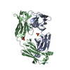


 PDBj
PDBj

 Assembly

 Mass: 18.015 Da / Num. of mol.: 664 / Source method: isolated from a natural source / Formula: H2O
 Sample preparation
 Processing