 Movie
Movie Controller


+ Open data

- Basic information
| Entry | Database: PDB / ID: 6mb8 | ||||||
|---|---|---|---|---|---|---|---|
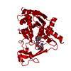
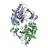
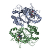
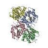
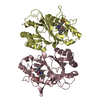
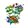
| Title | Apo structure of AAC-IIIb | ||||||
 Components Components | Aac(3)-IIIb protein | ||||||
 Keywords Keywords | TRANSFERASE/ANTIBIOTIC / acetyltransferase / promiscuity / GNAT / antibiotic resistance / ANTIBIOTIC / TRANSFERASE-ANTIBIOTIC complex | ||||||
| Function / homology | aminoglycoside 3-N-acetyltransferase activity / Aminoglycoside N(3)-acetyltransferase / Aminoglycoside 3-N-acetyltransferase / Aminoglycoside 3-N-acetyltransferase-like / Transferases; Acyltransferases; Transferring groups other than aminoacyl groups / response to antibiotic / Aminoglycoside N(3)-acetyltransferase Function and homology information Function and homology information | ||||||
| Biological species |   Pseudomonas aeruginosa (bacteria) Pseudomonas aeruginosa (bacteria) | ||||||
| Method |  X-RAY DIFFRACTION / MOLECULAR REPLACEMENT / Resolution: 1.6 Å X-RAY DIFFRACTION / MOLECULAR REPLACEMENT / Resolution: 1.6 Å | ||||||
 Authors Authors | Cuneo, M.J. / Kumar, P. | ||||||
 Citation Citation | Journal: J. Med. Chem. / Year: 2018 Title: Encoding of Promiscuity in an Aminoglycoside Acetyltransferase. Authors: Kumar, P. / Selvaraj, B. / Serpersu, E.H. / Cuneo, M.J. | ||||||
| History |
|
- Structure visualization
| Structure viewer | Molecule: MolmilJmol/JSmol |
|---|
- Downloads & links
-Download
| PDBx/mmCIF format | 6mb8.cif.gz | 136.3 KB | Display | PDBx/mmCIF format |
|---|---|---|---|---|
| PDB format | pdb6mb8.ent.gz | 103.2 KB | Display | PDB format |
| PDBx/mmJSON format | 6mb8.json.gz | Tree view | PDBx/mmJSON format | |
| Others |  Other downloads Other downloads |
-Validation report
| Arichive directory | https://data.pdbj.org/pub/pdb/validation_reports/mb/6mb8ftp://data.pdbj.org/pub/pdb/validation_reports/mb/6mb8 | HTTPS FTP |
|---|
-Related structure data
| Related structure data |  6mb4C 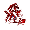 6mb5C  6mb6C  6mb7C  6mb9C  6bc3S S: Starting model for refinement C: citing same article ( |
|---|---|
| Similar structure data |


-Links
 PDBj
PDBj- Assembly
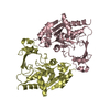
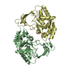
| Deposited unit | 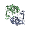
| ||||||||||||||||||||||||||||||||||||||||||||||||||||||||||||||||||||||||||||||||||||||||||
|---|---|---|---|---|---|---|---|---|---|---|---|---|---|---|---|---|---|---|---|---|---|---|---|---|---|---|---|---|---|---|---|---|---|---|---|---|---|---|---|---|---|---|---|---|---|---|---|---|---|---|---|---|---|---|---|---|---|---|---|---|---|---|---|---|---|---|---|---|---|---|---|---|---|---|---|---|---|---|---|---|---|---|---|---|---|---|---|---|---|---|---|
| 1 |
| ||||||||||||||||||||||||||||||||||||||||||||||||||||||||||||||||||||||||||||||||||||||||||
| 2 | 
| ||||||||||||||||||||||||||||||||||||||||||||||||||||||||||||||||||||||||||||||||||||||||||
| Unit cell |
| ||||||||||||||||||||||||||||||||||||||||||||||||||||||||||||||||||||||||||||||||||||||||||
| Components on special symmetry positions |
| ||||||||||||||||||||||||||||||||||||||||||||||||||||||||||||||||||||||||||||||||||||||||||
| Noncrystallographic symmetry (NCS) | NCS domain:
NCS domain segments: Ens-ID: 1
|
