 Movie
Movie Controller

[English] 日本語
 Yorodumi
- PDB-6m5b: X-ray crystal structure of cyclic-PIP and DNA complex in a revers... -
+ Open data

- Basic information
| Entry | Database: PDB / ID: 6m5b | |||||||||||||||||||||||||||||||
|---|---|---|---|---|---|---|---|---|---|---|---|---|---|---|---|---|---|---|---|---|---|---|---|---|---|---|---|---|---|---|---|---|
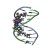
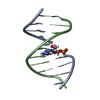
| Title | X-ray crystal structure of cyclic-PIP and DNA complex in a reverse binding orientation | |||||||||||||||||||||||||||||||
 Components Components | DNA (5'-D(* Keywords KeywordsDNA / PIP | Function / homology | Chem-S7E / DNA |  Function and homology information Function and homology informationBiological species | synthetic construct (others) | Method |  X-RAY DIFFRACTION / SYNCHROTRON / SAD / Resolution: 1.501 Å X-RAY DIFFRACTION / SYNCHROTRON / SAD / Resolution: 1.501 Å  Authors AuthorsAbe, K. / Hirose, Y. / Eki, H. / Takeda, K. / Bando, T. / Endo, M. / Sugiyama, H. | Funding support | |  Japan, 2items Japan, 2items
 CitationJournal: J.Am.Chem.Soc. / Year: 2020 CitationJournal: J.Am.Chem.Soc. / Year: 2020Title: X-ray Crystal Structure of a Cyclic-PIP-DNA Complex in the Reverse-Binding Orientation. Authors: Abe, K. / Hirose, Y. / Eki, H. / Takeda, K. / Bando, T. / Endo, M. / Sugiyama, H. History |
|
- Structure visualization
| Structure viewer | Molecule: MolmilJmol/JSmol |
|---|
- Downloads & links
-Download
| PDBx/mmCIF format | 6m5b.cif.gz | 23.5 KB | Display | PDBx/mmCIF format |
|---|---|---|---|---|
| PDB format | pdb6m5b.ent.gz | 12.7 KB | Display | PDB format |
| PDBx/mmJSON format | 6m5b.json.gz | Tree view | PDBx/mmJSON format | |
| Others |  Other downloads Other downloads |
-Validation report
| Arichive directory | https://data.pdbj.org/pub/pdb/validation_reports/m5/6m5bftp://data.pdbj.org/pub/pdb/validation_reports/m5/6m5b | HTTPS FTP |
|---|
-Related structure data
| Similar structure data |
|---|


-Links
 PDBj
PDBj

- Assembly
| Deposited unit | 
| |||||||||||||||
|---|---|---|---|---|---|---|---|---|---|---|---|---|---|---|---|---|
| 1 | 
| |||||||||||||||
| Unit cell |
| |||||||||||||||
| Components on special symmetry positions |
|
-Components
-DNA chain , 1 types, 1 molecules A
| #1: DNA chain | Mass: 3124.888 Da / Num. of mol.: 1 Source method: isolated from a genetically manipulated source Details: SF file contains Friedel pairs. / Source: (gene. exp.) synthetic construct (others) / Production host: synthetic construct (others) |
|---|
-Non-polymers , 5 types, 50 molecules 


| #2: Chemical | ChemComp-S7E /  Mass: 592.610 Da / Num. of mol.: 1 / Source method: obtained synthetically / Formula: C26H32N12O5 / Feature type: SUBJECT OF INVESTIGATION Mass: 592.610 Da / Num. of mol.: 1 / Source method: obtained synthetically / Formula: C26H32N12O5 / Feature type: SUBJECT OF INVESTIGATION |
|---|---|
| #3: Chemical | ChemComp-MG /  Mass: 24.305 Da / Num. of mol.: 1 / Source method: obtained synthetically / Formula: Mg Mass: 24.305 Da / Num. of mol.: 1 / Source method: obtained synthetically / Formula: Mg |
| #4: Chemical | ChemComp-NA /  Mass: 22.990 Da / Num. of mol.: 1 / Source method: obtained synthetically / Formula: Na Mass: 22.990 Da / Num. of mol.: 1 / Source method: obtained synthetically / Formula: Na |
| #5: Chemical | ChemComp-EDO /  Mass: 62.068 Da / Num. of mol.: 1 / Source method: obtained synthetically / Formula: C2H6O2 Mass: 62.068 Da / Num. of mol.: 1 / Source method: obtained synthetically / Formula: C2H6O2 |
| #6: Water | ChemComp-HOH / Mass: 18.015 Da / Num. of mol.: 46 / Source method: isolated from a natural source / Formula: H2O |
-Details
| Has ligand of interest | Y |
|---|
-Experimental details
-Experiment
| Experiment | Method: X-RAY DIFFRACTION / Number of used crystals: 1 |
|---|
- Sample preparation
| Crystal | Density Matthews: 3.71 Å3/Da / Density % sol: 66.83 % |
|---|---|
| Crystal grow | Temperature: 293 K / Method: vapor diffusion, hanging drop Details: 12.5% polyethylene glycol monomethyl ether 550, 2.5 mM magnesium chloride hexahydrate, 25 mM HEPES sodium, pH 7.0 |
-Data collection
| Diffraction | Mean temperature: 16.7 K / Serial crystal experiment: N | ||||||||||||||||||||||||||||||||||||||||||||||||||||||||||||||||||||||||||||||||||||||||||||||||||||
|---|---|---|---|---|---|---|---|---|---|---|---|---|---|---|---|---|---|---|---|---|---|---|---|---|---|---|---|---|---|---|---|---|---|---|---|---|---|---|---|---|---|---|---|---|---|---|---|---|---|---|---|---|---|---|---|---|---|---|---|---|---|---|---|---|---|---|---|---|---|---|---|---|---|---|---|---|---|---|---|---|---|---|---|---|---|---|---|---|---|---|---|---|---|---|---|---|---|---|---|---|---|
| Diffraction source | Source: SYNCHROTRON / Site: SPring-8 / Beamline: BL41XU / Wavelength: 0.91 Å | ||||||||||||||||||||||||||||||||||||||||||||||||||||||||||||||||||||||||||||||||||||||||||||||||||||
| Detector | Type: DECTRIS EIGER X 16M / Detector: PIXEL / Date: Oct 4, 2019 | ||||||||||||||||||||||||||||||||||||||||||||||||||||||||||||||||||||||||||||||||||||||||||||||||||||
| Radiation | Protocol: SINGLE WAVELENGTH / Monochromatic (M) / Laue (L): M / Scattering type: x-ray | ||||||||||||||||||||||||||||||||||||||||||||||||||||||||||||||||||||||||||||||||||||||||||||||||||||
| Radiation wavelength | Wavelength: 0.91 Å / Relative weight: 1 | ||||||||||||||||||||||||||||||||||||||||||||||||||||||||||||||||||||||||||||||||||||||||||||||||||||
| Reflection | Resolution: 1.5→41.118 Å / Num. obs: 14247 / % possible obs: 98.6 % / Redundancy: 14.328 % / Biso Wilson estimate: 23.982 Å2 / CC1/2: 1 / Rmerge(I) obs: 0.053 / Rrim(I) all: 0.054 / Χ2: 1.742 / Net I/σ(I): 26.37 / Num. measured all: 204128 | ||||||||||||||||||||||||||||||||||||||||||||||||||||||||||||||||||||||||||||||||||||||||||||||||||||
| Reflection shell | Diffraction-ID: 1
|
- Processing
| Software |
| ||||||||||||||||||||||||||||||||||||||||||||||||||||||
|---|---|---|---|---|---|---|---|---|---|---|---|---|---|---|---|---|---|---|---|---|---|---|---|---|---|---|---|---|---|---|---|---|---|---|---|---|---|---|---|---|---|---|---|---|---|---|---|---|---|---|---|---|---|---|---|
| Refinement | Method to determine structure: SAD / Resolution: 1.501→29.074 Å / SU ML: 0.17 / Cross valid method: THROUGHOUT / σ(F): 1.94 / Phase error: 21.75 / Stereochemistry target values: ML
| ||||||||||||||||||||||||||||||||||||||||||||||||||||||
| Solvent computation | Shrinkage radii: 0.9 Å / VDW probe radii: 1.11 Å / Solvent model: FLAT BULK SOLVENT MODEL | ||||||||||||||||||||||||||||||||||||||||||||||||||||||
| Displacement parameters | Biso max: 43.15 Å2 / Biso mean: 21.7689 Å2 / Biso min: 13.94 Å2 | ||||||||||||||||||||||||||||||||||||||||||||||||||||||
| Refinement step | Cycle: final / Resolution: 1.501→29.074 Å
| ||||||||||||||||||||||||||||||||||||||||||||||||||||||
| LS refinement shell | Refine-ID: X-RAY DIFFRACTION / Rfactor Rfree error: 0
|
