 Movie
Movie Controller


+ Open data

- Basic information
| Entry | Database: PDB / ID: 6m4b | ||||||
|---|---|---|---|---|---|---|---|
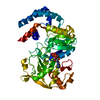
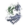
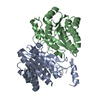
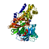
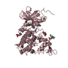
| Title | 1510-N membrane-bound stomatin-specific protease S97A mutant | ||||||
 Components Components | Membrane-bound protease PH1510 | ||||||
 Keywords Keywords | HYDROLASE / alpha/beta motif / protease / membrane protein stomatin | ||||||
| Function / homology |  Function and homology information Function and homology informationHydrolases; Acting on peptide bonds (peptidases); Serine endopeptidases / serine-type peptidase activity / proteolysis / membrane Similarity search - Function | ||||||
| Biological species |   Pyrococcus horikoshii (archaea) Pyrococcus horikoshii (archaea) | ||||||
| Method |  X-RAY DIFFRACTION / SYNCHROTRON / MOLECULAR REPLACEMENT / Resolution: 2.25 Å X-RAY DIFFRACTION / SYNCHROTRON / MOLECULAR REPLACEMENT / Resolution: 2.25 Å | ||||||
 Authors Authors | Yokoyama, H. / Suzuki, K. | ||||||
| Funding support |  Japan, 1items Japan, 1items
| ||||||
 Citation Citation | Journal: Acta Crystallogr.,Sect.D / Year: 2020 Title: Inactive dimeric structure of the protease domain of stomatin operon partner protein. Authors: Yokoyama, H. / Suzuki, K. / Hara, K. / Matsui, I. / Hashimoto, H. #1: Journal: J Synchrotron Radiat / Year: 2013Title: Structural and biochemical analysis of a thermostable membrane-bound stomatin-specific protease. Authors: Yokoyama, H. / Kobayashi, D. / Takizawa, N. / Fujii, S. / Matsui, I. | ||||||
| History |
|
- Structure visualization
| Structure viewer | Molecule: MolmilJmol/JSmol |
|---|
- Downloads & links
-Download
| PDBx/mmCIF format | 6m4b.cif.gz | 94.3 KB | Display | PDBx/mmCIF format |
|---|---|---|---|---|
| PDB format | pdb6m4b.ent.gz | 70.7 KB | Display | PDB format |
| PDBx/mmJSON format | 6m4b.json.gz | Tree view | PDBx/mmJSON format | |
| Others |  Other downloads Other downloads |
-Validation report
| Arichive directory | https://data.pdbj.org/pub/pdb/validation_reports/m4/6m4bftp://data.pdbj.org/pub/pdb/validation_reports/m4/6m4b | HTTPS FTP |
|---|
-Related structure data
| Related structure data |  3vivS S: Starting model for refinement |
|---|---|
| Similar structure data |


-Links
 PDBj
PDBj- Assembly
| Deposited unit | 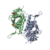
| ||||||||||||||||||
|---|---|---|---|---|---|---|---|---|---|---|---|---|---|---|---|---|---|---|---|
| 1 |
| ||||||||||||||||||
| Unit cell |
| ||||||||||||||||||
| Noncrystallographic symmetry (NCS) | NCS domain:
NCS domain segments: Component-ID: _ / Ens-ID: 1 / Beg auth comp-ID: LEU / Beg label comp-ID: LEU / End auth comp-ID: SER / End label comp-ID: SER / Refine code: _ / Auth seq-ID: 19 - 225 / Label seq-ID: 5 - 211
|
-Components
| #1: Protein | Mass: 25410.129 Da / Num. of mol.: 2 / Mutation: S97A Source method: isolated from a genetically manipulated source Source: (gene. exp.) Pyrococcus horikoshii (strain ATCC 700860 / DSM 12428 / JCM 9974 / NBRC 100139 / OT-3) (archaea)Strain: ATCC 700860 / DSM 12428 / JCM 9974 / NBRC 100139 / OT-3 Gene: PH1510 / Plasmid: pET21b / Production host:  References: UniProt: O59179, Hydrolases; Acting on peptide bonds (peptidases); Serine endopeptidases #2: Chemical |   Mass: 96.063 Da / Num. of mol.: 2 / Source method: obtained synthetically / Formula: SO4 / Feature type: SUBJECT OF INVESTIGATION Mass: 96.063 Da / Num. of mol.: 2 / Source method: obtained synthetically / Formula: SO4 / Feature type: SUBJECT OF INVESTIGATION#3: Water | ChemComp-HOH / |  Mass: 18.015 Da / Num. of mol.: 53 / Source method: isolated from a natural source / Formula: H2O Mass: 18.015 Da / Num. of mol.: 53 / Source method: isolated from a natural source / Formula: H2OHas ligand of interest | Y | |
|---|
-Experimental details
-Experiment
| Experiment | Method: X-RAY DIFFRACTION / Number of used crystals: 1 |
|---|
- Sample preparation
| Crystal | Density Matthews: 2.5 Å3/Da / Density % sol: 50.77 % |
|---|---|
| Crystal grow | Temperature: 293 K / Method: vapor diffusion, sitting drop / pH: 8 / Details: 0.18 M ammonium sulfate, 27% PEG4000 |
-Data collection
| Diffraction | Mean temperature: 100 K / Serial crystal experiment: N |
|---|---|
| Diffraction source | Source: SYNCHROTRON / Site: Photon Factory / Beamline: BL-17A / Wavelength: 0.98 Å |
| Detector | Type: ADSC QUANTUM 270 / Detector: CCD / Date: Jun 27, 2014 / Details: mirror |
| Radiation | Protocol: SINGLE WAVELENGTH / Monochromatic (M) / Laue (L): M / Scattering type: x-ray |
| Radiation wavelength | Wavelength: 0.98 Å / Relative weight: 1 |
| Reflection | Resolution: 2.25→20 Å / Num. obs: 24012 / % possible obs: 99.8 % / Redundancy: 11.3 % / Biso Wilson estimate: 42.8 Å2 / Rmerge(I) obs: 0.077 / Net I/σ(I): 21.9 |
| Reflection shell | Resolution: 2.25→2.37 Å / Rmerge(I) obs: 0.976 / Mean I/σ(I) obs: 2.8 / Num. unique obs: 3490 / % possible all: 100 |
- Processing
| Software |
| ||||||||||||||||||||||||||||||||||||||||||||||||||||||||||||
|---|---|---|---|---|---|---|---|---|---|---|---|---|---|---|---|---|---|---|---|---|---|---|---|---|---|---|---|---|---|---|---|---|---|---|---|---|---|---|---|---|---|---|---|---|---|---|---|---|---|---|---|---|---|---|---|---|---|---|---|---|---|
| Refinement | Method to determine structure: MOLECULAR REPLACEMENT Starting model: 3viv Resolution: 2.25→19.53 Å / Cor.coef. Fo:Fc: 0.96 / Cor.coef. Fo:Fc free: 0.939 / SU B: 6.167 / SU ML: 0.15 / Cross valid method: THROUGHOUT / σ(F): 0 / ESU R: 0.274 / ESU R Free: 0.211 Details: HYDROGENS HAVE BEEN ADDED IN THE RIDING POSITIONS U VALUES : REFINED INDIVIDUALLY
| ||||||||||||||||||||||||||||||||||||||||||||||||||||||||||||
| Solvent computation | Ion probe radii: 0.8 Å / Shrinkage radii: 0.8 Å / VDW probe radii: 1.2 Å | ||||||||||||||||||||||||||||||||||||||||||||||||||||||||||||
| Displacement parameters | Biso max: 150.07 Å2 / Biso mean: 52.99 Å2 / Biso min: 28.67 Å2
| ||||||||||||||||||||||||||||||||||||||||||||||||||||||||||||
| Refinement step | Cycle: final / Resolution: 2.25→19.53 Å
| ||||||||||||||||||||||||||||||||||||||||||||||||||||||||||||
| Refine LS restraints |
| ||||||||||||||||||||||||||||||||||||||||||||||||||||||||||||
| Refine LS restraints NCS | Ens-ID: 1 / Number: 25924 / Refine-ID: X-RAY DIFFRACTION / Type: interatomic distance / Rms dev position: 0.09 Å / Weight position: 0.05
| ||||||||||||||||||||||||||||||||||||||||||||||||||||||||||||
| LS refinement shell | Resolution: 2.25→2.308 Å / Rfactor Rfree error: 0 / Total num. of bins used: 20
|
