 Movie
Movie Controller

[English] 日本語
 Yorodumi
- PDB-6lx0: Structure of Leptospira santarosai serovar shermani LRR protein L... -
+ Open data

- Basic information
| Entry | Database: PDB / ID: 6lx0 | ||||||
|---|---|---|---|---|---|---|---|
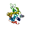
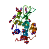
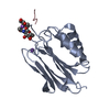
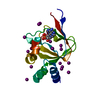
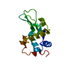
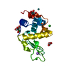
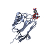
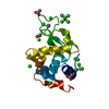
| Title | Structure of Leptospira santarosai serovar shermani LRR protein LSS11580 | ||||||
 Components Components | Membrane protein | ||||||
 Keywords Keywords | UNKNOWN FUNCTION / Leptospira / Leptospirosis / Leucine-rich repeat / CELL ADHESION / MEMBRANE PROTEIN | ||||||
| Function / homology | : / Leucine-rich repeats, bacterial type / Leucine rich repeat / Leucine-rich repeat, typical subtype / Leucine-rich repeats, typical (most populated) subfamily / Leucine-rich repeat profile. / Leucine-rich repeat / Leucine-rich repeat domain superfamily / Membrane protein Function and homology information Function and homology information | ||||||
| Biological species |  Leptospira santarosai serovar Shermani str. LT 821 (bacteria) Leptospira santarosai serovar Shermani str. LT 821 (bacteria) | ||||||
| Method |  X-RAY DIFFRACTION / SYNCHROTRON / MOLECULAR REPLACEMENT / Resolution: 1.99 Å X-RAY DIFFRACTION / SYNCHROTRON / MOLECULAR REPLACEMENT / Resolution: 1.99 Å | ||||||
 Authors Authors | Chu, C.H. / Hsu, S.H. / Yang, C.W. / Sun, Y.J. | ||||||
| Funding support |  Taiwan, 1items Taiwan, 1items
| ||||||
 Citation Citation | Journal: Biochem.J. / Year: 2020 Title: Crystal structure of Leptospira leucine-rich repeat 20 reveals a novel E-cadherin binding protein to induce NGAL expression in HK2 cells. Authors: Hsu, S.H. / Chu, C.H. / Tian, Y.C. / Chang, M.Y. / Chou, L.F. / Sun, Y.J. / Yang, C.W. | ||||||
| History |
|
- Structure visualization
| Structure viewer | Molecule: MolmilJmol/JSmol |
|---|
- Downloads & links
-Download
| PDBx/mmCIF format | 6lx0.cif.gz | 45.3 KB | Display | PDBx/mmCIF format |
|---|---|---|---|---|
| PDB format | pdb6lx0.ent.gz | 28.3 KB | Display | PDB format |
| PDBx/mmJSON format | 6lx0.json.gz | Tree view | PDBx/mmJSON format | |
| Others |  Other downloads Other downloads |
-Validation report
| Summary document | 6lx0_validation.pdf.gz | 427.5 KB | Display | wwPDB validaton report |
|---|---|---|---|---|
| Full document | 6lx0_full_validation.pdf.gz | 429.5 KB | Display | |
| Data in XML | 6lx0_validation.xml.gz | 7.8 KB | Display | |
| Data in CIF | 6lx0_validation.cif.gz | 10.1 KB | Display | |
| Arichive directory | https://data.pdbj.org/pub/pdb/validation_reports/lx/6lx0ftp://data.pdbj.org/pub/pdb/validation_reports/lx/6lx0 | HTTPS FTP |
-Related structure data
| Related structure data |  4pq8S S: Starting model for refinement |
|---|---|
| Similar structure data |

-Links
 PDBj
PDBj


- Assembly
| Deposited unit | 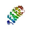
| ||||||||
|---|---|---|---|---|---|---|---|---|---|
| 1 |
| ||||||||
| Unit cell |
|
-Components
| #1: Protein | Mass: 24581.539 Da / Num. of mol.: 1 Source method: isolated from a genetically manipulated source Source: (gene. exp.) Leptospira santarosai serovar Shermani str. LT 821 (bacteria)Gene: LSS_11580 / Plasmid: pET30b / Production host: |
|---|---|
| #2: Water | ChemComp-HOH /  Mass: 18.015 Da / Num. of mol.: 77 / Source method: isolated from a natural source / Formula: H2O Mass: 18.015 Da / Num. of mol.: 77 / Source method: isolated from a natural source / Formula: H2O |
-Experimental details
-Experiment
| Experiment | Method: X-RAY DIFFRACTION / Number of used crystals: 1 |
|---|
- Sample preparation
| Crystal grow | Temperature: 289 K / Method: vapor diffusion, sitting drop / pH: 8.5 Details: 0.1M Tris hydrochloride (pH 8.5), 0.2M Sodium acetate trihydrate, 30% (w/v) Polyethylene glycol 4000 |
|---|
-Data collection
| Diffraction | Mean temperature: 100 K / Serial crystal experiment: N |
|---|---|
| Diffraction source | Source: SYNCHROTRON / Site: NSRRC / Beamline: BL15A1 / Wavelength: 1 Å |
| Detector | Type: MAR CCD 130 mm / Detector: CCD / Date: Oct 14, 2014 |
| Radiation | Protocol: SINGLE WAVELENGTH / Monochromatic (M) / Laue (L): M / Scattering type: x-ray |
| Radiation wavelength | Wavelength: 1 Å / Relative weight: 1 |
| Reflection | Resolution: 1.99→44.03 Å / Num. obs: 10011 / % possible obs: 99.7 % / Redundancy: 12.6 % / CC1/2: 0.998 / CC star: 0.999 / Net I/σ(I): 3.4 |
| Reflection shell | Resolution: 1.99→2.02 Å / Redundancy: 13.2 % / Num. unique obs: 486 / CC1/2: 0.896 / CC star: 0.972 / % possible all: 100 |
- Processing
| Software |
| ||||||||||||||||||||||||||||||
|---|---|---|---|---|---|---|---|---|---|---|---|---|---|---|---|---|---|---|---|---|---|---|---|---|---|---|---|---|---|---|---|
| Refinement | Method to determine structure: MOLECULAR REPLACEMENT Starting model: 4PQ8 Resolution: 1.99→19.959 Å / SU ML: 0.18 / Cross valid method: THROUGHOUT / σ(F): 1.36 / Phase error: 22.35 / Stereochemistry target values: ML
| ||||||||||||||||||||||||||||||
| Solvent computation | Shrinkage radii: 0.9 Å / VDW probe radii: 1.11 Å / Solvent model: FLAT BULK SOLVENT MODEL | ||||||||||||||||||||||||||||||
| Displacement parameters | Biso max: 61.37 Å2 / Biso mean: 25.1418 Å2 / Biso min: 10.5 Å2 | ||||||||||||||||||||||||||||||
| Refinement step | Cycle: final / Resolution: 1.99→19.959 Å
| ||||||||||||||||||||||||||||||
| Refine LS restraints |
| ||||||||||||||||||||||||||||||
| LS refinement shell | Refine-ID: X-RAY DIFFRACTION / Rfactor Rfree error: 0
|
