 Movie
Movie Controller

[English] 日本語
 Yorodumi
- PDB-6lwy: Crystal structure of Laterosporulin3, bacteriocin produced by Bre... -
+ Open data

- Basic information
| Entry | Database: PDB / ID: 6lwy | ||||||
|---|---|---|---|---|---|---|---|
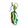
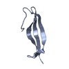
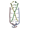
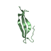
| Title | Crystal structure of Laterosporulin3, bacteriocin produced by Brevibacillus sp. strain SKR3 | ||||||
 Components Components | Laterosporulin3 | ||||||
 Keywords Keywords | TOXIN / Bacteriocin / Antimicrobial peptide / Laterosporulin / defensin | ||||||
| Function / homology | Laterosporulin defensin-like peptide / Laterosporulin defensin-like peptide / BROMIDE ION / Putative laterosporulin Function and homology information Function and homology information | ||||||
| Biological species |  Brevibacillus sp. SKR3 (bacteria) Brevibacillus sp. SKR3 (bacteria) | ||||||
| Method |  X-RAY DIFFRACTION / MOLECULAR REPLACEMENT / Resolution: 2.3 Å X-RAY DIFFRACTION / MOLECULAR REPLACEMENT / Resolution: 2.3 Å | ||||||
 Authors Authors | Thakur, K.G. / Solanki, V. | ||||||
| Funding support |  India, 1items India, 1items
| ||||||
 Citation Citation | Journal: To Be Published Title: Crystal structure of Laterosporulin3, bacteriocin produced by Brevibacillus sp. strain SKR3 Authors: Thakur, K.G. / Solanki, V. | ||||||
| History |
|
- Structure visualization
| Structure viewer | Molecule: MolmilJmol/JSmol |
|---|
- Downloads & links
-Download
| PDBx/mmCIF format | 6lwy.cif.gz | 34.4 KB | Display | PDBx/mmCIF format |
|---|---|---|---|---|
| PDB format | pdb6lwy.ent.gz | 21.9 KB | Display | PDB format |
| PDBx/mmJSON format | 6lwy.json.gz | Tree view | PDBx/mmJSON format | |
| Others |  Other downloads Other downloads |
-Validation report
| Arichive directory | https://data.pdbj.org/pub/pdb/validation_reports/lw/6lwyftp://data.pdbj.org/pub/pdb/validation_reports/lw/6lwy | HTTPS FTP |
|---|
-Related structure data
| Related structure data |  4ozkS S: Starting model for refinement |
|---|---|
| Similar structure data |
-Links
 PDBj
PDBj

- Assembly
| Deposited unit | 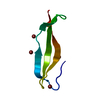
| ||||||||
|---|---|---|---|---|---|---|---|---|---|
| 1 |
| ||||||||
| Unit cell |
| ||||||||
| Components on special symmetry positions |
|
-Components
| #1: Protein/peptide | Mass: 5626.371 Da / Num. of mol.: 1 / Source method: isolated from a natural source / Source: (natural) Brevibacillus sp. SKR3 (bacteria) / Strain: SKR3 / References: UniProt: A0A075R4X6*PLUS | ||||||
|---|---|---|---|---|---|---|---|
| #2: Chemical | ChemComp-BR /   Mass: 79.904 Da / Num. of mol.: 4 / Source method: obtained synthetically / Formula: Br / Feature type: SUBJECT OF INVESTIGATION Mass: 79.904 Da / Num. of mol.: 4 / Source method: obtained synthetically / Formula: Br / Feature type: SUBJECT OF INVESTIGATION#3: Water | ChemComp-HOH / |  Mass: 18.015 Da / Num. of mol.: 4 / Source method: isolated from a natural source / Formula: H2O Mass: 18.015 Da / Num. of mol.: 4 / Source method: isolated from a natural source / Formula: H2OHas ligand of interest | Y | Has protein modification | Y | |
-Experimental details
-Experiment
| Experiment | Method: X-RAY DIFFRACTION / Number of used crystals: 1 |
|---|
- Sample preparation
| Crystal | Density Matthews: 1.89 Å3/Da / Density % sol: 35.01 % |
|---|---|
| Crystal grow | Temperature: 293 K / Method: vapor diffusion, sitting drop / pH: 5 Details: 0.2 M potassium bromide, 0.2 M potassium thiocyanate, 0.1 M sodium acetate, 3% w/v PGA-LM (Polyglycolic acid, 200-400 kDa) and 3% w/v PEG (Polyethylene glycol, 20 kDa), pH 5 |
-Data collection
| Diffraction | Mean temperature: 100 K / Serial crystal experiment: N | |||||||||||||||||||||||||||||||||
|---|---|---|---|---|---|---|---|---|---|---|---|---|---|---|---|---|---|---|---|---|---|---|---|---|---|---|---|---|---|---|---|---|---|---|
| Diffraction source | Source: ROTATING ANODE / Type: RIGAKU MICROMAX-007 HF / Wavelength: 1.5418 Å | |||||||||||||||||||||||||||||||||
| Detector | Type: MAR scanner 345 mm plate / Detector: IMAGE PLATE / Date: Nov 9, 2014 | |||||||||||||||||||||||||||||||||
| Radiation | Protocol: SINGLE WAVELENGTH / Monochromatic (M) / Laue (L): M / Scattering type: x-ray | |||||||||||||||||||||||||||||||||
| Radiation wavelength | Wavelength: 1.5418 Å / Relative weight: 1 | |||||||||||||||||||||||||||||||||
| Reflection | Resolution: 2.3→36.64 Å / Num. obs: 2214 / % possible obs: 100 % / Redundancy: 14 % / CC1/2: 0.999 / Rmerge(I) obs: 0.131 / Rpim(I) all: 0.037 / Rrim(I) all: 0.14 / Rsym value: 0.131 / Net I/σ(I): 16.6 | |||||||||||||||||||||||||||||||||
| Reflection shell | Diffraction-ID: 1
|
- Processing
| Software |
| ||||||||||||||||||||||||||||||||||||||||||||||||||||||||||||||||||||||||||||||||||||||||||||||||||||||||||||||||||||||||||||||||||||||||||||||||||||||
|---|---|---|---|---|---|---|---|---|---|---|---|---|---|---|---|---|---|---|---|---|---|---|---|---|---|---|---|---|---|---|---|---|---|---|---|---|---|---|---|---|---|---|---|---|---|---|---|---|---|---|---|---|---|---|---|---|---|---|---|---|---|---|---|---|---|---|---|---|---|---|---|---|---|---|---|---|---|---|---|---|---|---|---|---|---|---|---|---|---|---|---|---|---|---|---|---|---|---|---|---|---|---|---|---|---|---|---|---|---|---|---|---|---|---|---|---|---|---|---|---|---|---|---|---|---|---|---|---|---|---|---|---|---|---|---|---|---|---|---|---|---|---|---|---|---|---|---|---|---|---|---|
| Refinement | Method to determine structure: MOLECULAR REPLACEMENT Starting model: 4OZK Resolution: 2.3→36.639 Å / SU ML: 0.32 / Cross valid method: THROUGHOUT / σ(F): 1.36 / Phase error: 21.81
| ||||||||||||||||||||||||||||||||||||||||||||||||||||||||||||||||||||||||||||||||||||||||||||||||||||||||||||||||||||||||||||||||||||||||||||||||||||||
| Solvent computation | Shrinkage radii: 0.6 Å / VDW probe radii: 1 Å | ||||||||||||||||||||||||||||||||||||||||||||||||||||||||||||||||||||||||||||||||||||||||||||||||||||||||||||||||||||||||||||||||||||||||||||||||||||||
| Displacement parameters | Biso max: 128.61 Å2 / Biso mean: 35.2329 Å2 / Biso min: 13.88 Å2 | ||||||||||||||||||||||||||||||||||||||||||||||||||||||||||||||||||||||||||||||||||||||||||||||||||||||||||||||||||||||||||||||||||||||||||||||||||||||
| Refinement step | Cycle: final / Resolution: 2.3→36.639 Å
| ||||||||||||||||||||||||||||||||||||||||||||||||||||||||||||||||||||||||||||||||||||||||||||||||||||||||||||||||||||||||||||||||||||||||||||||||||||||
| LS refinement shell |
| ||||||||||||||||||||||||||||||||||||||||||||||||||||||||||||||||||||||||||||||||||||||||||||||||||||||||||||||||||||||||||||||||||||||||||||||||||||||
| Refinement TLS params. | Method: refined / Refine-ID: X-RAY DIFFRACTION
| ||||||||||||||||||||||||||||||||||||||||||||||||||||||||||||||||||||||||||||||||||||||||||||||||||||||||||||||||||||||||||||||||||||||||||||||||||||||
| Refinement TLS group |
|
