 Movie
Movie Controller

[English] 日本語
 Yorodumi
- PDB-6ll9: Crystal structure of D-alanine-D-alanine ligase from Aeromonas hy... -
+ Open data

- Basic information
| Entry | Database: PDB / ID: 6ll9 | ||||||
|---|---|---|---|---|---|---|---|
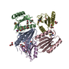
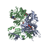
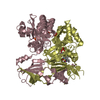
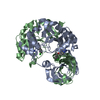
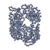
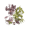
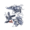
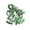
| Title | Crystal structure of D-alanine-D-alanine ligase from Aeromonas hydrophila | ||||||
 Components Components | D-alanine--D-alanine ligase | ||||||
 Keywords Keywords | LIGASE / Aeromonas hydrophila / D-alanine-D-alanine ligase / cell wall | ||||||
| Function / homology |  Function and homology information Function and homology informationD-alanine-D-alanine ligase / D-alanine-D-alanine ligase activity / peptidoglycan biosynthetic process / cell wall organization / regulation of cell shape / ATP binding / metal ion binding / cytoplasm / cytosol Similarity search - Function | ||||||
| Biological species |  Aeromonas hydrophila (bacteria) Aeromonas hydrophila (bacteria) | ||||||
| Method |  X-RAY DIFFRACTION / SYNCHROTRON / MOLECULAR REPLACEMENT / Resolution: 2.7 Å X-RAY DIFFRACTION / SYNCHROTRON / MOLECULAR REPLACEMENT / Resolution: 2.7 Å | ||||||
 Authors Authors | Zhang, H. | ||||||
 Citation Citation | Journal: J.Agric.Food Chem. / Year: 2020 Title: Insights into the Inhibition ofAeromonas hydrophilad-Alanine-d-Alanine Ligase by Integration of Kinetics and Structural Analysis. Authors: Zhang, Y. / Gong, S. / Wang, X. / Muhammad, M. / Li, Y. / Meng, S. / Li, Q. / Liu, D. / Zhang, H. | ||||||
| History |
|
- Structure visualization
| Structure viewer | Molecule: MolmilJmol/JSmol |
|---|
- Downloads & links
-Download
| PDBx/mmCIF format | 6ll9.cif.gz | 231.9 KB | Display | PDBx/mmCIF format |
|---|---|---|---|---|
| PDB format | pdb6ll9.ent.gz | 184.1 KB | Display | PDB format |
| PDBx/mmJSON format | 6ll9.json.gz | Tree view | PDBx/mmJSON format | |
| Others |  Other downloads Other downloads |
-Validation report
| Arichive directory | https://data.pdbj.org/pub/pdb/validation_reports/ll/6ll9ftp://data.pdbj.org/pub/pdb/validation_reports/ll/6ll9 | HTTPS FTP |
|---|
-Related structure data
| Related structure data | 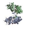 6dgiS S: Starting model for refinement |
|---|---|
| Similar structure data |


-Links
 PDBj
PDBj


- Assembly
| Deposited unit | 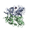
| |||||||||||||||||||||||||||||||||||||||||||||||||||||||||||||||||||||||||||||||||||||||||||||
|---|---|---|---|---|---|---|---|---|---|---|---|---|---|---|---|---|---|---|---|---|---|---|---|---|---|---|---|---|---|---|---|---|---|---|---|---|---|---|---|---|---|---|---|---|---|---|---|---|---|---|---|---|---|---|---|---|---|---|---|---|---|---|---|---|---|---|---|---|---|---|---|---|---|---|---|---|---|---|---|---|---|---|---|---|---|---|---|---|---|---|---|---|---|---|
| 1 |
| |||||||||||||||||||||||||||||||||||||||||||||||||||||||||||||||||||||||||||||||||||||||||||||
| Unit cell |
| |||||||||||||||||||||||||||||||||||||||||||||||||||||||||||||||||||||||||||||||||||||||||||||
| Noncrystallographic symmetry (NCS) | NCS domain:
NCS domain segments: Ens-ID: 1
|