 Movie
Movie Controller

[English] 日本語
 Yorodumi
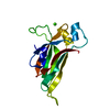
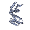
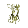
- PDB-6lk3: The Functional Characterization and Crystal Structure of Type II ... -
+ Open data

- Basic information
| Entry | Database: PDB / ID: 6lk3 | ||||||
|---|---|---|---|---|---|---|---|
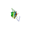
| Title | The Functional Characterization and Crystal Structure of Type II Peptidyl Carrier Protein ColA1a in Collismycins Biosynthesis | ||||||
 Components Components | Putative free-standing acyl carrier protein | ||||||
 Keywords Keywords | BIOSYNTHETIC PROTEIN / Biosynthesis / ColA1a / Collismycins / NRPS / Peptidyl Carrier Protein | ||||||
| Function / homology | ACP-like superfamily / Putative free-standing acyl carrier protein Function and homology information Function and homology information | ||||||
| Biological species |  Streptomyces sp. CS40 (bacteria) Streptomyces sp. CS40 (bacteria) | ||||||
| Method |  X-RAY DIFFRACTION / SYNCHROTRON / SAD / Resolution: 2.1 Å X-RAY DIFFRACTION / SYNCHROTRON / SAD / Resolution: 2.1 Å | ||||||
 Authors Authors | Ma, X.Y. / Wang, G.Y. / Liu, T. / Chi, C.B. / Zhang, Z.Y. / Yang, D.H. / Liu, W. / Ma, M. | ||||||
| Funding support |  China, 1items China, 1items
| ||||||
 Citation Citation | Journal: Chin.J.Chem. / Year: 2020 Title: The functional characterization and crystal structure of type II peptidyl carrier protein ColA1a in collismycins biosynthesis. Authors: Ma, X.Y. / Wang, G.Y. / Liu, T. / Chi, C.B. / Zhang, Z.Y. / Yang, D.H. / Liu, W. / Ma, M. | ||||||
| History |
|
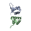
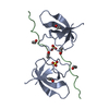
- Structure visualization
| Structure viewer | Molecule: MolmilJmol/JSmol |
|---|
- Downloads & links
-Download
| PDBx/mmCIF format | 6lk3.cif.gz | 45.8 KB | Display | PDBx/mmCIF format |
|---|---|---|---|---|
| PDB format | pdb6lk3.ent.gz | 31 KB | Display | PDB format |
| PDBx/mmJSON format | 6lk3.json.gz | Tree view | PDBx/mmJSON format | |
| Others |  Other downloads Other downloads |
-Validation report
| Arichive directory | https://data.pdbj.org/pub/pdb/validation_reports/lk/6lk3ftp://data.pdbj.org/pub/pdb/validation_reports/lk/6lk3 | HTTPS FTP |
|---|
-Related structure data
| Similar structure data |
|---|

-Links
 PDBj
PDBj
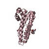
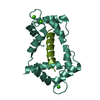
- Assembly
| Deposited unit | 
| ||||||||
|---|---|---|---|---|---|---|---|---|---|
| 1 |
| ||||||||
| Unit cell |
|
-Components
| #1: Protein | Mass: 11883.458 Da / Num. of mol.: 2 Source method: isolated from a genetically manipulated source Source: (gene. exp.) Streptomyces sp. CS40 (bacteria) / Gene: clmP / Production host: #2: Water | ChemComp-HOH / |  Mass: 18.015 Da / Num. of mol.: 101 / Source method: isolated from a natural source / Formula: H2O Mass: 18.015 Da / Num. of mol.: 101 / Source method: isolated from a natural source / Formula: H2OSequence details | The source organism is Streptomyces roseosporus NRRL 11379. So, There is no genome sequence of ...The source organism is Streptomyces roseosporus NRRL 11379. So, There is no genome sequence of Streptomyces roseosporus NRRL 11379 in the database. | |
|---|
-Experimental details
-Experiment
| Experiment | Method: X-RAY DIFFRACTION / Number of used crystals: 1 |
|---|
- Sample preparation
| Crystal | Density Matthews: 1.78 Å3/Da / Density % sol: 30.87 % |
|---|---|
| Crystal grow | Temperature: 289 K / Method: vapor diffusion, hanging drop Details: 0.2 M sodium chloride, 0.1 M Bis-Tris pH 6.25, 25% w/v polyethylene glycol 3,350 |
-Data collection
| Diffraction | Mean temperature: 100 K / Serial crystal experiment: N |
|---|---|
| Diffraction source | Source: SYNCHROTRON / Site: SSRF / Beamline: BL17U1 / Wavelength: 0.97917 Å |
| Detector | Type: DECTRIS EIGER X 16M / Detector: PIXEL / Date: May 13, 2018 |
| Radiation | Protocol: SINGLE WAVELENGTH / Monochromatic (M) / Laue (L): M / Scattering type: x-ray |
| Radiation wavelength | Wavelength: 0.97917 Å / Relative weight: 1 |
| Reflection | Resolution: 2.1→39.51 Å / Num. obs: 9816 / % possible obs: 98.1 % / Redundancy: 13.3 % / Rmerge(I) obs: 0.139 / Net I/σ(I): 4.6 |
| Reflection shell | Resolution: 2.1→2.18 Å / Rmerge(I) obs: 0.473 / Num. unique obs: 503 |
- Processing
| Software |
| ||||||||||||||||||||||||||||||||||||||||||||||||||||||||||||
|---|---|---|---|---|---|---|---|---|---|---|---|---|---|---|---|---|---|---|---|---|---|---|---|---|---|---|---|---|---|---|---|---|---|---|---|---|---|---|---|---|---|---|---|---|---|---|---|---|---|---|---|---|---|---|---|---|---|---|---|---|---|
| Refinement | Method to determine structure: SAD / Resolution: 2.1→39.51 Å / Cor.coef. Fo:Fc: 0.963 / Cor.coef. Fo:Fc free: 0.937 / SU B: 3.852 / SU ML: 0.105 / Cross valid method: THROUGHOUT / σ(F): 0 / ESU R: 0.186 / ESU R Free: 0.165 / Stereochemistry target values: MAXIMUM LIKELIHOOD Details: HYDROGENS HAVE BEEN ADDED IN THE RIDING POSITIONS U VALUES : REFINED INDIVIDUALLY
| ||||||||||||||||||||||||||||||||||||||||||||||||||||||||||||
| Solvent computation | Ion probe radii: 0.8 Å / Shrinkage radii: 0.8 Å / VDW probe radii: 1.2 Å / Solvent model: MASK | ||||||||||||||||||||||||||||||||||||||||||||||||||||||||||||
| Displacement parameters | Biso max: 77.45 Å2 / Biso mean: 24.982 Å2 / Biso min: 9.64 Å2
| ||||||||||||||||||||||||||||||||||||||||||||||||||||||||||||
| Refinement step | Cycle: final / Resolution: 2.1→39.51 Å
| ||||||||||||||||||||||||||||||||||||||||||||||||||||||||||||
| Refine LS restraints |
| ||||||||||||||||||||||||||||||||||||||||||||||||||||||||||||
| LS refinement shell | Resolution: 2.1→2.15 Å / Rfactor Rfree error: 0
|
