Resolution: 1.95→42.43 Å / Cor.coef. Fo:Fc: 0.946 / Cor.coef. Fo:Fc free: 0.904 / SU B: 5.423 / SU ML: 0.147 / Cross valid method: THROUGHOUT / ESU R: 0.203 / ESU R Free: 0.185 / Stereochemistry target values: MAXIMUM LIKELIHOOD / Details: HYDROGENS HAVE BEEN USED IF PRESENT IN THE INPUT
Rfactor
Num. reflection
% reflection
Selection details
Rfree
0.25025
1358
9.9 %
RANDOM
Rwork
0.18731
-
-
-
obs
0.19339
12398
99.99 %
-
Solvent computation
Ion probe radii: 0.8 Å / Shrinkage radii: 0.8 Å / VDW probe radii: 1.2 Å / Solvent model: MASK
 Movie
Movie Controller

 Yorodumi
 Open data
 Basic information
 Components
 Keywords
 Function and homology information
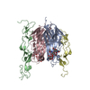
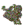
 Bombyx mori cytoplasmic polyhedrosis virus
 X-RAY DIFFRACTION /
  Authors
 Japan, 1items
  Citation
 Structure visualization
 Downloads & links
 Other downloads
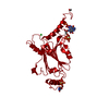


 PDBj
PDBj Assembly


 Spodoptera frugiperda (fall armyworm) / References: UniProt: P11041
 Mass: 18.015 Da / Num. of mol.: 52 / Source method: isolated from a natural source / Formula: H2O
 Sample preparation
 Processing