 Movie
Movie Controller

[English] 日本語
 Yorodumi
- PDB-6lcl: TtGalA, alpha-galactosidase from Thermus thermophilus in complex ... -
+ Open data

- Basic information
| Entry | Database: PDB / ID: 6lcl | |||||||||
|---|---|---|---|---|---|---|---|---|---|---|
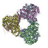
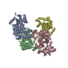
| Title | TtGalA, alpha-galactosidase from Thermus thermophilus in complex with stachyose | |||||||||
 Components Components | Alpha-galactosidase | |||||||||
 Keywords Keywords | HYDROLASE / alpha-galactosidase / hexamer assembly / substrate specificity / thermostable / stachyose | |||||||||
| Function / homology |  Function and homology information Function and homology information | |||||||||
| Biological species |   Thermus thermophilus (bacteria) Thermus thermophilus (bacteria) | |||||||||
| Method |  X-RAY DIFFRACTION / SYNCHROTRON / MOLECULAR REPLACEMENT / Resolution: 3.2 Å X-RAY DIFFRACTION / SYNCHROTRON / MOLECULAR REPLACEMENT / Resolution: 3.2 Å | |||||||||
 Authors Authors | Chen, S.C. / Hsu, C.H. | |||||||||
| Funding support |  Taiwan, 2items Taiwan, 2items
| |||||||||
 Citation Citation | Journal: J.Agric.Food Chem. / Year: 2020 Title: Crystal Structure of alpha-Galactosidase from Thermus thermophilus : Insight into Hexamer Assembly and Substrate Specificity. Authors: Chen, S.C. / Wu, S.P. / Chang, Y.Y. / Hwang, T.S. / Lee, T.H. / Hsu, C.H. | |||||||||
| History |
|
- Structure visualization
| Structure viewer | Molecule: MolmilJmol/JSmol |
|---|
- Downloads & links
-Download
| PDBx/mmCIF format | 6lcl.cif.gz | 350.5 KB | Display | PDBx/mmCIF format |
|---|---|---|---|---|
| PDB format | pdb6lcl.ent.gz | 229.7 KB | Display | PDB format |
| PDBx/mmJSON format | 6lcl.json.gz | Tree view | PDBx/mmJSON format | |
| Others |  Other downloads Other downloads |
-Validation report
| Arichive directory | https://data.pdbj.org/pub/pdb/validation_reports/lc/6lclftp://data.pdbj.org/pub/pdb/validation_reports/lc/6lcl | HTTPS FTP |
|---|
-Related structure data


-Links
 PDBj
PDBj- Assembly
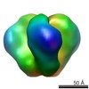
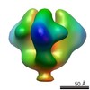
| Deposited unit | 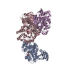
| ||||||||||||
|---|---|---|---|---|---|---|---|---|---|---|---|---|---|
| 1 | 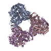
| ||||||||||||
| Unit cell |
|
-Components
| #1: Protein | Mass: 53986.309 Da / Num. of mol.: 3 Source method: isolated from a genetically manipulated source Source: (gene. exp.) Thermus thermophilus (strain HB8 / ATCC 27634 / DSM 579) (bacteria)Gene: TTHB115 / Production host: #2: Polysaccharide | Source method: isolated from a genetically manipulated source Has ligand of interest | Y | |
|---|
-Experimental details
-Experiment
| Experiment | Method: X-RAY DIFFRACTION / Number of used crystals: 1 |
|---|
- Sample preparation
| Crystal | Density Matthews: 3.06 Å3/Da / Density % sol: 59.83 % |
|---|---|
| Crystal grow | Temperature: 283 K / Method: vapor diffusion, sitting drop / Details: 0.1M CAPSO pH 9.2, 0.2M Li2SO4, 10% PEG 3000 |
-Data collection
| Diffraction | Mean temperature: 100 K / Serial crystal experiment: N |
|---|---|
| Diffraction source | Source: SYNCHROTRON / Site: NSRRC / Beamline: BL13C1 / Wavelength: 0.9762 Å |
| Detector | Type: ADSC QUANTUM 315r / Detector: CCD / Date: Nov 10, 2012 |
| Radiation | Protocol: SINGLE WAVELENGTH / Monochromatic (M) / Laue (L): M / Scattering type: x-ray |
| Radiation wavelength | Wavelength: 0.9762 Å / Relative weight: 1 |
| Reflection | Resolution: 3.2→50 Å / Num. obs: 32852 / % possible obs: 99.3 % / Redundancy: 7 % / Biso Wilson estimate: 62.78 Å2 / Rmerge(I) obs: 0.112 / Net I/σ(I): 19.2 |
| Reflection shell | Resolution: 3.2→3.31 Å / Rmerge(I) obs: 0.504 / Num. unique obs: 3187 |
- Processing
| Software |
| |||||||||||||||||||||||||||||||||||||||||||||||||||||||||||||||||||||||||||||||||||||||||||||||||||||||||
|---|---|---|---|---|---|---|---|---|---|---|---|---|---|---|---|---|---|---|---|---|---|---|---|---|---|---|---|---|---|---|---|---|---|---|---|---|---|---|---|---|---|---|---|---|---|---|---|---|---|---|---|---|---|---|---|---|---|---|---|---|---|---|---|---|---|---|---|---|---|---|---|---|---|---|---|---|---|---|---|---|---|---|---|---|---|---|---|---|---|---|---|---|---|---|---|---|---|---|---|---|---|---|---|---|---|---|
| Refinement | Method to determine structure: MOLECULAR REPLACEMENT Starting model: apo structure Resolution: 3.2→25.22 Å / SU ML: 0.3173 / Cross valid method: FREE R-VALUE / σ(F): 1.35 / Phase error: 20.4208
| |||||||||||||||||||||||||||||||||||||||||||||||||||||||||||||||||||||||||||||||||||||||||||||||||||||||||
| Solvent computation | Shrinkage radii: 0.9 Å / VDW probe radii: 1.11 Å | |||||||||||||||||||||||||||||||||||||||||||||||||||||||||||||||||||||||||||||||||||||||||||||||||||||||||
| Displacement parameters | Biso mean: 53.87 Å2 | |||||||||||||||||||||||||||||||||||||||||||||||||||||||||||||||||||||||||||||||||||||||||||||||||||||||||
| Refinement step | Cycle: LAST / Resolution: 3.2→25.22 Å
| |||||||||||||||||||||||||||||||||||||||||||||||||||||||||||||||||||||||||||||||||||||||||||||||||||||||||
| Refine LS restraints |
| |||||||||||||||||||||||||||||||||||||||||||||||||||||||||||||||||||||||||||||||||||||||||||||||||||||||||
| LS refinement shell |
|
