 Movie
Movie Controller

[English] 日本語
 Yorodumi
- PDB-6lac: The structural basis of the beta-carbonic anhydrase CafD (C39A mu... -
+ Open data

- Basic information
| Entry | Database: PDB / ID: 6lac | |||||||||
|---|---|---|---|---|---|---|---|---|---|---|
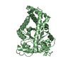
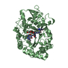
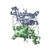
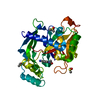
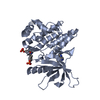
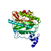
| Title | The structural basis of the beta-carbonic anhydrase CafD (C39A mutant) of the filamentous fungus Aspergillus fumigatus | |||||||||
 Components Components | Carbonic anhydrase | |||||||||
 Keywords Keywords | LYASE / beta-class carbonic anhydrase / Aspergillus fumigatus / CafD | |||||||||
| Function / homology |  Function and homology information Function and homology informationcellular response to carbon dioxide / carbonic anhydrase / carbonate dehydratase activity / cellular response to oxidative stress / zinc ion binding / cytoplasm Similarity search - Function | |||||||||
| Biological species |  | |||||||||
| Method |  X-RAY DIFFRACTION / SYNCHROTRON / MOLECULAR REPLACEMENT / Resolution: 1.3 Å X-RAY DIFFRACTION / SYNCHROTRON / MOLECULAR REPLACEMENT / Resolution: 1.3 Å | |||||||||
 Authors Authors | Jiin, M.S. / Kim, S. / Kim, N.J. / Hong, S. / Kim, S. / Sung, J. | |||||||||
| Funding support |  Korea, Republic Of, 2items Korea, Republic Of, 2items
| |||||||||
 Citation Citation | Journal: To be published Title: The structural analysis of non-catalytic zinc binding site in the minor beta-carbonic anhydrase CafD Authors: Jin, M.S. / Kim, S. / Kim, N.J. / Hong, S. / Kim, S. / Sung, J. | |||||||||
| History |
|
- Structure visualization
| Structure viewer | Molecule: MolmilJmol/JSmol |
|---|
- Downloads & links
-Download
| PDBx/mmCIF format | 6lac.cif.gz | 83.5 KB | Display | PDBx/mmCIF format |
|---|---|---|---|---|
| PDB format | pdb6lac.ent.gz | 61.4 KB | Display | PDB format |
| PDBx/mmJSON format | 6lac.json.gz | Tree view | PDBx/mmJSON format | |
| Others |  Other downloads Other downloads |
-Validation report
| Summary document | 6lac_validation.pdf.gz | 1.5 MB | Display | wwPDB validaton report |
|---|---|---|---|---|
| Full document | 6lac_full_validation.pdf.gz | 1.5 MB | Display | |
| Data in XML | 6lac_validation.xml.gz | 15.7 KB | Display | |
| Data in CIF | 6lac_validation.cif.gz | 22.1 KB | Display | |
| Arichive directory | https://data.pdbj.org/pub/pdb/validation_reports/la/6lacftp://data.pdbj.org/pub/pdb/validation_reports/la/6lac | HTTPS FTP |
-Related structure data
| Related structure data |  6laiC  6jqeS S: Starting model for refinement C: citing same article ( |
|---|---|
| Similar structure data |


-Links
 PDBj
PDBj
- Assembly
| Deposited unit | 
| ||||||||
|---|---|---|---|---|---|---|---|---|---|
| 1 |
| ||||||||
| Unit cell |
|
-Components
| #1: Protein | Mass: 20197.867 Da / Num. of mol.: 2 / Mutation: C39A Source method: isolated from a genetically manipulated source Source: (gene. exp.) Strain: ATCC MYA-4609 / Af293 / CBS 101355 / FGSC A1100 / Gene: AFUA_8G06554 / Production host:  #2: Chemical |   Mass: 65.409 Da / Num. of mol.: 2 / Source method: isolated from a natural source / Formula: Zn / Feature type: SUBJECT OF INVESTIGATION Mass: 65.409 Da / Num. of mol.: 2 / Source method: isolated from a natural source / Formula: Zn / Feature type: SUBJECT OF INVESTIGATION#3: Water | ChemComp-HOH / |  Mass: 18.015 Da / Num. of mol.: 153 / Source method: isolated from a natural source / Formula: H2O Mass: 18.015 Da / Num. of mol.: 153 / Source method: isolated from a natural source / Formula: H2OHas ligand of interest | Y | |
|---|
-Experimental details
-Experiment
| Experiment | Method: X-RAY DIFFRACTION / Number of used crystals: 1 |
|---|
- Sample preparation
| Crystal | Density Matthews: 2.15 Å3/Da / Density % sol: 42.68 % |
|---|---|
| Crystal grow | Temperature: 295 K / Method: vapor diffusion, sitting drop Details: 28 % PEG3350, 200 mM MgCl2, 100 mM Tris-HCl pH 6.0-9.5 |
-Data collection
| Diffraction | Mean temperature: 77 K / Serial crystal experiment: N |
|---|---|
| Diffraction source | Source: SYNCHROTRON / Site: PAL/PLS / Beamline: 11C / Wavelength: 0.979 Å |
| Detector | Type: DECTRIS PILATUS3 S 6M / Detector: PIXEL / Date: Jun 1, 2019 |
| Radiation | Protocol: SINGLE WAVELENGTH / Monochromatic (M) / Laue (L): M / Scattering type: x-ray |
| Radiation wavelength | Wavelength: 0.979 Å / Relative weight: 1 |
| Reflection | Resolution: 1.3→50 Å / Num. obs: 75211 / % possible obs: 99.6 % / Redundancy: 3.4 % / Rsym value: 0.02 / Net I/σ(I): 30.3 |
| Reflection shell | Resolution: 1.3→1.35 Å / Num. unique obs: 7904 / Rsym value: 0.2 |
- Processing
| Software |
| ||||||||||||||||||||||||||||||||||||||||||||||||||||||||||||
|---|---|---|---|---|---|---|---|---|---|---|---|---|---|---|---|---|---|---|---|---|---|---|---|---|---|---|---|---|---|---|---|---|---|---|---|---|---|---|---|---|---|---|---|---|---|---|---|---|---|---|---|---|---|---|---|---|---|---|---|---|---|
| Refinement | Method to determine structure: MOLECULAR REPLACEMENT Starting model: 6JQE Resolution: 1.3→33.92 Å / Cor.coef. Fo:Fc: 0.964 / Cor.coef. Fo:Fc free: 0.957 / SU B: 0.846 / SU ML: 0.036 / Cross valid method: THROUGHOUT / σ(F): 0 / ESU R: 0.053 / ESU R Free: 0.054 / Stereochemistry target values: MAXIMUM LIKELIHOOD Details: HYDROGENS HAVE BEEN ADDED IN THE RIDING POSITIONS U VALUES : REFINED INDIVIDUALLY
| ||||||||||||||||||||||||||||||||||||||||||||||||||||||||||||
| Solvent computation | Ion probe radii: 0.8 Å / Shrinkage radii: 0.8 Å / VDW probe radii: 1.2 Å / Solvent model: MASK | ||||||||||||||||||||||||||||||||||||||||||||||||||||||||||||
| Displacement parameters | Biso max: 72.18 Å2 / Biso mean: 19.216 Å2 / Biso min: 8.1 Å2
| ||||||||||||||||||||||||||||||||||||||||||||||||||||||||||||
| Refinement step | Cycle: final / Resolution: 1.3→33.92 Å
| ||||||||||||||||||||||||||||||||||||||||||||||||||||||||||||
| Refine LS restraints |
| ||||||||||||||||||||||||||||||||||||||||||||||||||||||||||||
| LS refinement shell | Resolution: 1.3→1.333 Å / Rfactor Rfree error: 0 / Total num. of bins used: 20
|
