 Movie
Movie Controller


+ Open data

- Basic information
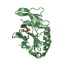
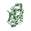
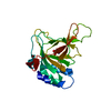
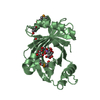
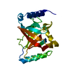
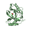
| Entry | Database: PDB / ID: 6kyc | ||||||
|---|---|---|---|---|---|---|---|
| Title | Structure of the S207A mutant of Clostridium difficile sortase B | ||||||
 Components Components | Putative peptidase C60B, sortase B | ||||||
 Keywords Keywords | HYDROLASE / sortase B / cysteine transpeptidase / Clostridium difficile | ||||||
| Function / homology |  Function and homology information Function and homology information | ||||||
| Biological species |  Peptoclostridium difficile 630 (bacteria) Peptoclostridium difficile 630 (bacteria) | ||||||
| Method |  X-RAY DIFFRACTION / SYNCHROTRON / MOLECULAR REPLACEMENT / Resolution: 2.604 Å X-RAY DIFFRACTION / SYNCHROTRON / MOLECULAR REPLACEMENT / Resolution: 2.604 Å | ||||||
 Authors Authors | Kang, C.Y. / Huang, I.H. / Wu, T.Y. / Chang, J.C. / Hsiao, Y.Y. / Cheng, C.H. / Tsai, W.J. / Hsu, K.C. / Wang, S.Y. | ||||||
| Funding support |  Taiwan, 1items Taiwan, 1items
| ||||||
 Citation Citation | Journal: J.Biol.Chem. / Year: 2020 Title: Functional analysis ofClostridium difficilesortase B reveals key residues for catalytic activity and substrate specificity. Authors: Kang, C.Y. / Huang, I.H. / Chou, C.C. / Wu, T.Y. / Chang, J.C. / Hsiao, Y.Y. / Cheng, C.H. / Tsai, W.J. / Hsu, K.C. / Wang, S. | ||||||
| History |
|
- Structure visualization
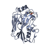
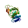
| Structure viewer | Molecule: MolmilJmol/JSmol |
|---|
- Downloads & links
-Download
| PDBx/mmCIF format | 6kyc.cif.gz | 91.8 KB | Display | PDBx/mmCIF format |
|---|---|---|---|---|
| PDB format | pdb6kyc.ent.gz | 68 KB | Display | PDB format |
| PDBx/mmJSON format | 6kyc.json.gz | Tree view | PDBx/mmJSON format | |
| Others |  Other downloads Other downloads |
-Validation report
| Arichive directory | https://data.pdbj.org/pub/pdb/validation_reports/ky/6kycftp://data.pdbj.org/pub/pdb/validation_reports/ky/6kyc | HTTPS FTP |
|---|
-Related structure data
| Related structure data |  6kydC 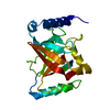 5gyjS S: Starting model for refinement C: citing same article ( |
|---|---|
| Similar structure data |
-Links
 PDBj
PDBj- Assembly
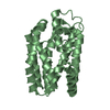
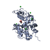
| Deposited unit | 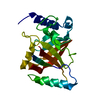
| ||||||||
|---|---|---|---|---|---|---|---|---|---|
| 1 |
| ||||||||
| Unit cell |
|
-Components
| #1: Protein | Mass: 26173.283 Da / Num. of mol.: 1 / Mutation: S207A Source method: isolated from a genetically manipulated source Source: (gene. exp.) Peptoclostridium difficile 630 (bacteria)Strain: 630 / Gene: CD630_27180 / Production host: |
|---|---|
| #2: Water | ChemComp-HOH /  Mass: 18.015 Da / Num. of mol.: 24 / Source method: isolated from a natural source / Formula: H2O Mass: 18.015 Da / Num. of mol.: 24 / Source method: isolated from a natural source / Formula: H2O |
-Experimental details
-Experiment
| Experiment | Method: X-RAY DIFFRACTION / Number of used crystals: 1 |
|---|
- Sample preparation
| Crystal | Density Matthews: 2.82 Å3/Da / Density % sol: 56.35 % |
|---|---|
| Crystal grow | Temperature: 295.15 K / Method: vapor diffusion, hanging drop / pH: 4.2 / Details: 23% PEG3350, 0.1M Glycine, 0.1M citric acid(pH4.2) / PH range: 4.0-4.2 |
-Data collection
| Diffraction | Mean temperature: 100 K / Serial crystal experiment: N | |||||||||||||||||||||||||||||||||||||||||||||||||||||||||||||||||||||||||||||||||||||||||||||||||||
|---|---|---|---|---|---|---|---|---|---|---|---|---|---|---|---|---|---|---|---|---|---|---|---|---|---|---|---|---|---|---|---|---|---|---|---|---|---|---|---|---|---|---|---|---|---|---|---|---|---|---|---|---|---|---|---|---|---|---|---|---|---|---|---|---|---|---|---|---|---|---|---|---|---|---|---|---|---|---|---|---|---|---|---|---|---|---|---|---|---|---|---|---|---|---|---|---|---|---|---|---|
| Diffraction source | Source: SYNCHROTRON / Site: NSRRC / Beamline: BL13B1 / Wavelength: 1 Å | |||||||||||||||||||||||||||||||||||||||||||||||||||||||||||||||||||||||||||||||||||||||||||||||||||
| Detector | Type: ADSC QUANTUM 315r / Detector: CCD / Date: Oct 24, 2018 | |||||||||||||||||||||||||||||||||||||||||||||||||||||||||||||||||||||||||||||||||||||||||||||||||||
| Radiation | Protocol: SINGLE WAVELENGTH / Monochromatic (M) / Laue (L): M / Scattering type: x-ray | |||||||||||||||||||||||||||||||||||||||||||||||||||||||||||||||||||||||||||||||||||||||||||||||||||
| Radiation wavelength | Wavelength: 1 Å / Relative weight: 1 | |||||||||||||||||||||||||||||||||||||||||||||||||||||||||||||||||||||||||||||||||||||||||||||||||||
| Reflection | Resolution: 2.6→30 Å / Num. obs: 9127 / % possible obs: 99.4 % / Redundancy: 7.1 % / Biso Wilson estimate: 41.32 Å2 / Rmerge(I) obs: 0.052 / Rpim(I) all: 0.02 / Rrim(I) all: 0.056 / Χ2: 0.937 / Net I/σ(I): 15.1 / Num. measured all: 64443 | |||||||||||||||||||||||||||||||||||||||||||||||||||||||||||||||||||||||||||||||||||||||||||||||||||
| Reflection shell | Diffraction-ID: 1
|
- Processing
| Software |
| ||||||||||||||||||||||||||||||||||||||||
|---|---|---|---|---|---|---|---|---|---|---|---|---|---|---|---|---|---|---|---|---|---|---|---|---|---|---|---|---|---|---|---|---|---|---|---|---|---|---|---|---|---|
| Refinement | Method to determine structure: MOLECULAR REPLACEMENT Starting model: 5GYJ Resolution: 2.604→28.513 Å / SU ML: 0.31 / Cross valid method: THROUGHOUT / σ(F): 1.38 / Phase error: 24.49 / Stereochemistry target values: ML
| ||||||||||||||||||||||||||||||||||||||||
| Solvent computation | Shrinkage radii: 0.9 Å / VDW probe radii: 1.11 Å / Solvent model: FLAT BULK SOLVENT MODEL | ||||||||||||||||||||||||||||||||||||||||
| Displacement parameters | Biso max: 98.41 Å2 / Biso mean: 44.4123 Å2 / Biso min: 16.96 Å2 | ||||||||||||||||||||||||||||||||||||||||
| Refinement step | Cycle: final / Resolution: 2.604→28.513 Å
| ||||||||||||||||||||||||||||||||||||||||
| Refine LS restraints |
| ||||||||||||||||||||||||||||||||||||||||
| LS refinement shell | Refine-ID: X-RAY DIFFRACTION / Rfactor Rfree error: 0
| ||||||||||||||||||||||||||||||||||||||||
| Refinement TLS params. | Method: refined / Origin x: -8.9583 Å / Origin y: -34.8259 Å / Origin z: -42.0221 Å
| ||||||||||||||||||||||||||||||||||||||||
| Refinement TLS group |
|
