 Movie
Movie Controller

[English] 日本語
 Yorodumi
- PDB-6krf: An X-ray structure of ferric F43Y/F46S sperm whale myoglobin in c... -
+ Open data

- Basic information
| Entry | Database: PDB / ID: 6krf | ||||||
|---|---|---|---|---|---|---|---|
| Title | An X-ray structure of ferric F43Y/F46S sperm whale myoglobin in complex with guaiacol | ||||||
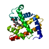
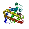
 Components Components | Myoglobin | ||||||
 Keywords Keywords | OXYGEN STORAGE / myoglobin | ||||||
| Function / homology |  Function and homology information Function and homology informationOxidoreductases; Acting on other nitrogenous compounds as donors / nitrite reductase activity / sarcoplasm / Oxidoreductases; Acting on a peroxide as acceptor; Peroxidases / removal of superoxide radicals / oxygen carrier activity / peroxidase activity / oxygen binding / heme binding / extracellular exosome / metal ion binding Similarity search - Function | ||||||
| Biological species |  | ||||||
| Method |  X-RAY DIFFRACTION / SYNCHROTRON / MOLECULAR REPLACEMENT / Resolution: 1.86 Å X-RAY DIFFRACTION / SYNCHROTRON / MOLECULAR REPLACEMENT / Resolution: 1.86 Å | ||||||
 Authors Authors | Yuan, H. / Lin, Y.W. | ||||||
 Citation Citation | Journal: Acs Catalysis / Year: 2020 Title: A Catalytic Binding Site Together with a Distal Tyr in MyoglobinAffords Catalytic Efficiencies Similar to Natural Peroxidases. Authors: Zhang, P. / Yuan, H. / Xu, J. / Wang, X.J. / Gao, S.Q. / Tan, X. / Lin, Y.W. | ||||||
| History |
|
- Structure visualization
| Structure viewer | Molecule: MolmilJmol/JSmol |
|---|
- Downloads & links
-Download
| PDBx/mmCIF format | 6krf.cif.gz | 61.3 KB | Display | PDBx/mmCIF format |
|---|---|---|---|---|
| PDB format | pdb6krf.ent.gz | 34.6 KB | Display | PDB format |
| PDBx/mmJSON format | 6krf.json.gz | Tree view | PDBx/mmJSON format | |
| Others |  Other downloads Other downloads |
-Validation report
| Arichive directory | https://data.pdbj.org/pub/pdb/validation_reports/kr/6krfftp://data.pdbj.org/pub/pdb/validation_reports/kr/6krf | HTTPS FTP |
|---|
-Related structure data
| Related structure data |  6krcC  5iksS S: Starting model for refinement C: citing same article ( |
|---|---|
| Similar structure data |


-Links
 PDBj
PDBj


- Assembly
| Deposited unit | 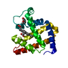
| ||||||||||||
|---|---|---|---|---|---|---|---|---|---|---|---|---|---|
| 1 |
| ||||||||||||
| Unit cell |
|
-Components
| #1: Protein | Mass: 17190.855 Da / Num. of mol.: 1 / Mutation: F43Y, F46S Source method: isolated from a genetically manipulated source Source: (gene. exp.)  |
|---|---|
| #2: Chemical | ChemComp-HEM /   Mass: 616.487 Da / Num. of mol.: 1 / Source method: obtained synthetically / Formula: C34H32FeN4O4 Mass: 616.487 Da / Num. of mol.: 1 / Source method: obtained synthetically / Formula: C34H32FeN4O4 |
| #3: Chemical | ChemComp-JZ3 / 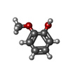  Mass: 124.137 Da / Num. of mol.: 1 / Source method: obtained synthetically / Formula: C7H8O2 / Feature type: SUBJECT OF INVESTIGATION Mass: 124.137 Da / Num. of mol.: 1 / Source method: obtained synthetically / Formula: C7H8O2 / Feature type: SUBJECT OF INVESTIGATION |
| #4: Water | ChemComp-HOH /  Mass: 18.015 Da / Num. of mol.: 197 / Source method: isolated from a natural source / Formula: H2O Mass: 18.015 Da / Num. of mol.: 197 / Source method: isolated from a natural source / Formula: H2O |
| Has ligand of interest | Y |
-Experimental details
-Experiment
| Experiment | Method: X-RAY DIFFRACTION / Number of used crystals: 1 |
|---|
- Sample preparation
| Crystal | Density Matthews: 2.21 Å3/Da / Density % sol: 44.31 % |
|---|---|
| Crystal grow | Temperature: 293 K / Method: vapor diffusion, hanging drop Details: 0.2 M Sodium acetate trihydrate, 0.1 M Sodium cacodylate trihydrate pH 6.5, 30% w/v Polyethylene glycol 8,000 |
-Data collection
| Diffraction | Mean temperature: 100 K / Serial crystal experiment: N |
|---|---|
| Diffraction source | Source: SYNCHROTRON / Site: SSRF  / Beamline: BL18U1 / Wavelength: 0.98 Å / Beamline: BL18U1 / Wavelength: 0.98 Å |
| Detector | Type: DECTRIS PILATUS3 S 6M / Detector: PIXEL / Date: Jun 15, 2018 |
| Radiation | Protocol: SINGLE WAVELENGTH / Monochromatic (M) / Laue (L): M / Scattering type: x-ray |
| Radiation wavelength | Wavelength: 0.98 Å / Relative weight: 1 |
| Reflection | Resolution: 1.86→50 Å / Num. obs: 13245 / % possible obs: 99.8 % / Redundancy: 5.8 % / Biso Wilson estimate: 24.3 Å2 / Rmerge(I) obs: 0.116 / Rpim(I) all: 0.048 / Net I/σ(I): 19.21 |
| Reflection shell | Resolution: 1.86→1.91 Å / Rmerge(I) obs: 0.793 / Mean I/σ(I) obs: 2.19 / Num. unique obs: 884 / Rpim(I) all: 0.427 |
- Processing
| Software |
| ||||||||||||||||||||||||||||||||||||||||||
|---|---|---|---|---|---|---|---|---|---|---|---|---|---|---|---|---|---|---|---|---|---|---|---|---|---|---|---|---|---|---|---|---|---|---|---|---|---|---|---|---|---|---|---|
| Refinement | Method to determine structure: MOLECULAR REPLACEMENT Starting model: 5iks Resolution: 1.86→30.78 Å / SU ML: 0.1773 / Cross valid method: FREE R-VALUE / σ(F): 1.34 / Phase error: 22.2587
| ||||||||||||||||||||||||||||||||||||||||||
| Solvent computation | Shrinkage radii: 0.9 Å / VDW probe radii: 1.11 Å | ||||||||||||||||||||||||||||||||||||||||||
| Displacement parameters | Biso mean: 28.64 Å2 | ||||||||||||||||||||||||||||||||||||||||||
| Refinement step | Cycle: LAST / Resolution: 1.86→30.78 Å
| ||||||||||||||||||||||||||||||||||||||||||
| Refine LS restraints |
| ||||||||||||||||||||||||||||||||||||||||||
| LS refinement shell |
|
