 Movie
Movie Controller


+ Open data

- Basic information
| Entry | Database: PDB / ID: 6k5u | ||||||
|---|---|---|---|---|---|---|---|
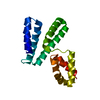
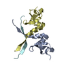
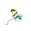
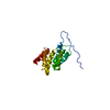
| Title | Crystal structure of the myb domain of S. pombe Tbf1 | ||||||
 Components Components | Telomeric DNA-binding factor trf1 | ||||||
 Keywords Keywords | DNA BINDING PROTEIN / Telomere binding protein | ||||||
| Function / homology |  Function and homology information Function and homology informationchromosome, telomeric repeat region / telomere maintenance via telomere lengthening / double-stranded telomeric DNA binding / telomere maintenance / chromatin / protein homodimerization activity / nucleus Similarity search - Function | ||||||
| Biological species |  | ||||||
| Method |  X-RAY DIFFRACTION / SYNCHROTRON / SAD / Resolution: 2.079 Å X-RAY DIFFRACTION / SYNCHROTRON / SAD / Resolution: 2.079 Å | ||||||
 Authors Authors | Zhou, Y.Z. / Wang, N.N. / Zhao, Y.C. / Zeng, Z.X. | ||||||
 Citation Citation | Journal: To Be Published Title: Crystal structure of the myb domain of S. pombe Tbf1 Authors: Zhou, Y.Z. / Wang, N.N. / Zhao, Y.C. / Zeng, Z.X. | ||||||
| History |
|
- Structure visualization
| Structure viewer | Molecule: MolmilJmol/JSmol |
|---|
- Downloads & links
-Download
| PDBx/mmCIF format | 6k5u.cif.gz | 46.5 KB | Display | PDBx/mmCIF format |
|---|---|---|---|---|
| PDB format | pdb6k5u.ent.gz | 32.4 KB | Display | PDB format |
| PDBx/mmJSON format | 6k5u.json.gz | Tree view | PDBx/mmJSON format | |
| Others |  Other downloads Other downloads |
-Validation report
| Arichive directory | https://data.pdbj.org/pub/pdb/validation_reports/k5/6k5uftp://data.pdbj.org/pub/pdb/validation_reports/k5/6k5u | HTTPS FTP |
|---|
-Related structure data
| Related structure data | |
|---|---|
| Similar structure data |


-Links
 PDBj
PDBj
- Assembly
| Deposited unit | 
| ||||||||
|---|---|---|---|---|---|---|---|---|---|
| 1 | 
| ||||||||
| 2 | 
| ||||||||
| Unit cell |
| ||||||||
| Components on special symmetry positions |
|
-Components
| #1: Protein | Mass: 9116.227 Da / Num. of mol.: 2 / Mutation: L420M Source method: isolated from a genetically manipulated source Source: (gene. exp.) Strain: 972 / ATCC 24843 / Gene: trf1, SPBC19G7.13 / Variant: 972 / ATCC 24843 / Production host:  #2: Water | ChemComp-HOH / |  Mass: 18.015 Da / Num. of mol.: 119 / Source method: isolated from a natural source / Formula: H2O Mass: 18.015 Da / Num. of mol.: 119 / Source method: isolated from a natural source / Formula: H2OHas protein modification | Y | Sequence details | L420M is mutated and MET is modified to MSE. | |
|---|
-Experimental details
-Experiment
| Experiment | Method: X-RAY DIFFRACTION / Number of used crystals: 1 |
|---|
- Sample preparation
| Crystal | Density Matthews: 3.17 Å3/Da / Density % sol: 61.14 % |
|---|---|
| Crystal grow | Temperature: 277.15 K / Method: vapor diffusion Details: 16% PEG 8000, 40mM Potassium phosphate dibasic, 20% Glycerol |
-Data collection
| Diffraction | Mean temperature: 100 K / Serial crystal experiment: N |
|---|---|
| Diffraction source | Source: SYNCHROTRON / Site: SSRF  / Beamline: BL18U1 / Wavelength: 0.97778 Å / Beamline: BL18U1 / Wavelength: 0.97778 Å |
| Detector | Type: DECTRIS EIGER X 16M / Detector: PIXEL / Date: Jan 15, 2017 |
| Radiation | Protocol: SINGLE WAVELENGTH / Monochromatic (M) / Laue (L): M / Scattering type: x-ray |
| Radiation wavelength | Wavelength: 0.97778 Å / Relative weight: 1 |
| Reflection | Resolution: 2.079→40 Å / Num. obs: 14514 / % possible obs: 100 % / Redundancy: 24.7 % / Biso Wilson estimate: 26.39 Å2 / Rmerge(I) obs: 0.185 / Rpim(I) all: 0.038 / Rrim(I) all: 0.189 / Χ2: 1.368 / Net I/σ(I): 32.455 |
| Reflection shell | Resolution: 2.1→2.14 Å / Redundancy: 23.4 % / Rmerge(I) obs: 0.873 / Num. unique obs: 696 / CC1/2: 0.964 / Rpim(I) all: 0.183 / Rrim(I) all: 0.893 / Χ2: 0.494 / % possible all: 100 |
-Phasing
| Phasing | Method: SAD |
|---|
- Processing
| Software |
| ||||||||||||||||||||||||||||||||||||||||||
|---|---|---|---|---|---|---|---|---|---|---|---|---|---|---|---|---|---|---|---|---|---|---|---|---|---|---|---|---|---|---|---|---|---|---|---|---|---|---|---|---|---|---|---|
| Refinement | Method to determine structure: SAD / Resolution: 2.079→37.262 Å / SU ML: 0.18 / Cross valid method: THROUGHOUT / σ(F): 1.37 / Phase error: 23.07
| ||||||||||||||||||||||||||||||||||||||||||
| Solvent computation | Shrinkage radii: 0.9 Å / VDW probe radii: 1.11 Å | ||||||||||||||||||||||||||||||||||||||||||
| Displacement parameters | Biso max: 63.07 Å2 / Biso mean: 29.0306 Å2 / Biso min: 13.44 Å2 | ||||||||||||||||||||||||||||||||||||||||||
| Refinement step | Cycle: final / Resolution: 2.079→37.262 Å
| ||||||||||||||||||||||||||||||||||||||||||
| LS refinement shell | Refine-ID: X-RAY DIFFRACTION / Rfactor Rfree error: 0 / Total num. of bins used: 5
|
