 Movie
Movie Controller


+ Open data

- Basic information
| Entry | Database: PDB / ID: 6k5p | ||||||
|---|---|---|---|---|---|---|---|
| Title | Structure of mosquito-larvicidal Binary toxin receptor, Cqm1 | ||||||
 Components Components | Binary toxin receptor protein | ||||||
 Keywords Keywords | PROTEIN BINDING / Amylomaltase / GH13_17 subfamily / Receptor for BinAB toxin / Cqm1 | ||||||
| Function / homology |  Function and homology information Function and homology informationalpha-glucosidase / carbohydrate metabolic process / membrane / metal ion binding Similarity search - Function | ||||||
| Biological species |  Culex quinquefasciatus (southern house mosquito) Culex quinquefasciatus (southern house mosquito) | ||||||
| Method |  X-RAY DIFFRACTION / SYNCHROTRON / MOLECULAR REPLACEMENT / Resolution: 1.805 Å X-RAY DIFFRACTION / SYNCHROTRON / MOLECULAR REPLACEMENT / Resolution: 1.805 Å | ||||||
 Authors Authors | Kumar, V. / Sharma, M. | ||||||
 Citation Citation | Journal: Int.J.Biol.Macromol. / Year: 2019 Title: Crystal structure of BinAB toxin receptor (Cqm1) protein and molecular dynamics simulations reveal the role of unique Ca(II) ion. Authors: Sharma, M. / Kumar, V. #1: Journal: Acta Crystallogr F Struct Biol Commun / Year: 2018Title: Mosquito-larvicidal binary toxin receptor protein (Cqm1): crystallization and X-ray crystallographic analysis. Authors: Sharma, M. / Lakshmi, A. / Gupta, G.D. / Kumar, V. #2: Journal: Insect Biochem. Mol. Biol. / Year: 2018Title: Receptor protein of Lysinibacillus sphaericus mosquito-larvicidal toxin displays amylomaltase activity. Authors: Sharma, M. / Gupta, G.D. / Kumar, V. | ||||||
| History |
|
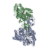
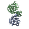
- Structure visualization
| Structure viewer | Molecule: MolmilJmol/JSmol |
|---|
- Downloads & links
-Download
| PDBx/mmCIF format | 6k5p.cif.gz | 939.6 KB | Display | PDBx/mmCIF format |
|---|---|---|---|---|
| PDB format | pdb6k5p.ent.gz | 776.7 KB | Display | PDB format |
| PDBx/mmJSON format | 6k5p.json.gz | Tree view | PDBx/mmJSON format | |
| Others |  Other downloads Other downloads |
-Validation report
| Arichive directory | https://data.pdbj.org/pub/pdb/validation_reports/k5/6k5pftp://data.pdbj.org/pub/pdb/validation_reports/k5/6k5p | HTTPS FTP |
|---|
-Related structure data
| Related structure data |  3wy1S S: Starting model for refinement |
|---|---|
| Similar structure data |


-Links
 PDBj
PDBj


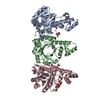
- Assembly
| Deposited unit | 
| ||||||||
|---|---|---|---|---|---|---|---|---|---|
| 1 | 
| ||||||||
| 2 | 
| ||||||||
| 3 |
| ||||||||
| Unit cell |
| ||||||||
| Details | Authors state that the protein 6K5P does not exist as tetramer, |
-Components
-Protein , 1 types, 4 molecules ABCD
| #1: Protein | Mass: 64744.902 Da / Num. of mol.: 4 Source method: isolated from a genetically manipulated source Source: (gene. exp.) Culex quinquefasciatus (southern house mosquito)Gene: Cqm1 / Production host:  |
|---|
-Non-polymers , 6 types, 2333 molecules 


| #2: Chemical | ChemComp-MG /  Mass: 24.305 Da / Num. of mol.: 4 Mass: 24.305 Da / Num. of mol.: 4Source method: isolated from a genetically manipulated source Formula: Mg #3: Chemical | ChemComp-CD /  Mass: 112.411 Da / Num. of mol.: 14 Mass: 112.411 Da / Num. of mol.: 14Source method: isolated from a genetically manipulated source Formula: Cd #4: Chemical | ChemComp-CA /  Mass: 40.078 Da / Num. of mol.: 4 / Source method: isolated from a natural source / Formula: Ca / Feature type: SUBJECT OF INVESTIGATION Mass: 40.078 Da / Num. of mol.: 4 / Source method: isolated from a natural source / Formula: Ca / Feature type: SUBJECT OF INVESTIGATION#5: Chemical | ChemComp-ACT /  Mass: 59.044 Da / Num. of mol.: 5 / Source method: obtained synthetically / Formula: C2H3O2 Mass: 59.044 Da / Num. of mol.: 5 / Source method: obtained synthetically / Formula: C2H3O2#6: Chemical |  Mass: 58.933 Da / Num. of mol.: 2 / Source method: obtained synthetically / Formula: Co Mass: 58.933 Da / Num. of mol.: 2 / Source method: obtained synthetically / Formula: Co#7: Water | ChemComp-HOH / | Mass: 18.015 Da / Num. of mol.: 2304 / Source method: isolated from a natural source / Formula: H2O |
|---|
-Details
| Sequence details | THE SEQUENCE OF THIS PROTEIN WAS NOT AVAILABLE AT THE UNIPROT KNOWLEDGEBASE DATABASE (UNIPROTKB) AT ...THE SEQUENCE OF THIS PROTEIN WAS NOT AVAILABLE AT THE UNIPROT KNOWLEDGEB |
|---|
-Experimental details
-Experiment
| Experiment | Method: X-RAY DIFFRACTION / Number of used crystals: 1 |
|---|
- Sample preparation
| Crystal | Density Matthews: 2.24 Å3/Da / Density % sol: 48.18 % |
|---|---|
| Crystal grow | Temperature: 295 K / Method: vapor diffusion, sitting drop / pH: 7.5 Details: 5mM CoCl2, 5mM CdCl2, 5mM MgCl2, 5mM NiCl2, 0.1mM HEPES pH 7.5, 12% PEG 3350 |
-Data collection
| Diffraction | Mean temperature: 100 K / Serial crystal experiment: N |
|---|---|
| Diffraction source | Source: SYNCHROTRON / Site: RRCAT INDUS-2  / Beamline: PX-BL21 / Wavelength: 0.9795 Å / Beamline: PX-BL21 / Wavelength: 0.9795 Å |
| Detector | Type: MAR scanner 345 mm plate / Detector: IMAGE PLATE / Date: Oct 3, 2018 |
| Radiation | Monochromator: Double Crystal Monochromator / Protocol: SINGLE WAVELENGTH / Monochromatic (M) / Laue (L): M / Scattering type: x-ray |
| Radiation wavelength | Wavelength: 0.9795 Å / Relative weight: 1 |
| Reflection | Resolution: 1.805→39.23 Å / Num. obs: 201511 / % possible obs: 93.9 % / Redundancy: 7.1 % / CC1/2: 0.998 / Rmerge(I) obs: 0.072 / Rpim(I) all: 0.044 / Rrim(I) all: 0.09 / Net I/σ(I): 14.4 |
| Reflection shell | Resolution: 1.805→1.9 Å / Redundancy: 7.3 % / Rmerge(I) obs: 0.532 / Mean I/σ(I) obs: 2.8 / Num. unique obs: 31009 / CC1/2: 0.929 / Rpim(I) all: 0.318 / Rrim(I) all: 0.621 / % possible all: 99.9 |
- Processing
| Software |
| ||||||||||||||||||||||||||||||||||||||||
|---|---|---|---|---|---|---|---|---|---|---|---|---|---|---|---|---|---|---|---|---|---|---|---|---|---|---|---|---|---|---|---|---|---|---|---|---|---|---|---|---|---|
| Refinement | Method to determine structure: MOLECULAR REPLACEMENT Starting model: 3WY1 Resolution: 1.805→29.828 Å / SU ML: 0.2 / Cross valid method: FREE R-VALUE / σ(F): 1.34 / Phase error: 24.38 Details: Metal-ligand distance were not restrained. SF FILE CONTAINS FRIEDEL PAIRS UNDER I/F_MINUS AND I/F_PLUS COLUMNS. Data at 1.9A was used for obtaining initial phases by MRSAD method. High ...Details: Metal-ligand distance were not restrained. SF FILE CONTAINS FRIEDEL PAIRS UNDER I/F_MINUS AND I/F_PLUS COLUMNS. Data at 1.9A was used for obtaining initial phases by MRSAD method. High resolution data acquired at 0.9795A was used for structure refinement.
| ||||||||||||||||||||||||||||||||||||||||
| Solvent computation | Shrinkage radii: 0.9 Å / VDW probe radii: 1.11 Å | ||||||||||||||||||||||||||||||||||||||||
| Refinement step | Cycle: LAST / Resolution: 1.805→29.828 Å
| ||||||||||||||||||||||||||||||||||||||||
| Refine LS restraints |
| ||||||||||||||||||||||||||||||||||||||||
| LS refinement shell | Resolution: 1.805→1.823 Å
| ||||||||||||||||||||||||||||||||||||||||
| Refinement TLS params. | Method: refined / Origin x: -17.6037 Å / Origin y: -1.0327 Å / Origin z: 74.8901 Å
| ||||||||||||||||||||||||||||||||||||||||
| Refinement TLS group | Selection details: all |
