 Movie
Movie Controller


+ Open data

- Basic information
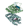
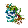
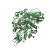
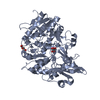
| Entry | Database: PDB / ID: 6k4d | |||||||||
|---|---|---|---|---|---|---|---|---|---|---|
| Title | Ancestral luciferase AncLamp in complex with ATP and D-luciferin | |||||||||
 Components Components | Ancestral luciferase AncLamp | |||||||||
 Keywords Keywords | OXIDOREDUCTASE / Luciferase / Bioluminescence / Ancestral protein / Molecular evolution | |||||||||
| Function / homology | Chem-D4F / Chem-ESJ Function and homology information Function and homology information | |||||||||
| Biological species |  Lampyridae (fireflies) Lampyridae (fireflies) | |||||||||
| Method |  X-RAY DIFFRACTION / SYNCHROTRON / MOLECULAR REPLACEMENT / Resolution: 1.7 Å X-RAY DIFFRACTION / SYNCHROTRON / MOLECULAR REPLACEMENT / Resolution: 1.7 Å | |||||||||
 Authors Authors | Oba, Y. / Konishi, K. / Yano, D. / Kato, D. / Shirai, T. | |||||||||
| Funding support |  Japan, 2items Japan, 2items
| |||||||||
 Citation Citation | Journal: Sci Adv / Year: 2020 Title: Resurrecting the ancient glow of the fireflies. Authors: Oba, Y. / Konishi, K. / Yano, D. / Shibata, H. / Kato, D. / Shirai, T. | |||||||||
| History |
|
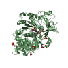
- Structure visualization
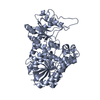
| Structure viewer | Molecule: MolmilJmol/JSmol |
|---|
- Downloads & links
-Download
| PDBx/mmCIF format | 6k4d.cif.gz | 140.5 KB | Display | PDBx/mmCIF format |
|---|---|---|---|---|
| PDB format | pdb6k4d.ent.gz | 105.5 KB | Display | PDB format |
| PDBx/mmJSON format | 6k4d.json.gz | Tree view | PDBx/mmJSON format | |
| Others |  Other downloads Other downloads |
-Validation report
| Arichive directory | https://data.pdbj.org/pub/pdb/validation_reports/k4/6k4dftp://data.pdbj.org/pub/pdb/validation_reports/k4/6k4d | HTTPS FTP |
|---|
-Related structure data
| Related structure data | 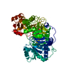 6k4cC 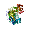 2d1rS S: Starting model for refinement C: citing same article ( |
|---|---|
| Similar structure data |


-Links
 PDBj
PDBj
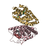
- Assembly
| Deposited unit | 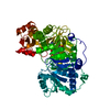
| ||||||||
|---|---|---|---|---|---|---|---|---|---|
| 1 |
| ||||||||
| Unit cell |
| ||||||||
| Components on special symmetry positions |
|
-Components
| #1: Protein | Mass: 60374.371 Da / Num. of mol.: 1 Source method: isolated from a genetically manipulated source Source: (gene. exp.) Lampyridae (fireflies) / Production host:  |
|---|---|
| #2: Chemical | ChemComp-D4F / [[(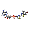  Mass: 609.529 Da / Num. of mol.: 1 / Source method: isolated from a natural source / Formula: C21H20N7O9PS2 / Feature type: SUBJECT OF INVESTIGATION Mass: 609.529 Da / Num. of mol.: 1 / Source method: isolated from a natural source / Formula: C21H20N7O9PS2 / Feature type: SUBJECT OF INVESTIGATION |
| #3: Chemical | ChemComp-ESJ / (  Mass: 280.323 Da / Num. of mol.: 1 Mass: 280.323 Da / Num. of mol.: 1Source method: isolated from a genetically manipulated source Formula: C11H8N2O3S2 / Feature type: SUBJECT OF INVESTIGATION |
| #4: Water | ChemComp-HOH /  Mass: 18.015 Da / Num. of mol.: 730 / Source method: isolated from a natural source / Formula: H2O Mass: 18.015 Da / Num. of mol.: 730 / Source method: isolated from a natural source / Formula: H2O |
| Has ligand of interest | Y |
-Experimental details
-Experiment
| Experiment | Method: X-RAY DIFFRACTION / Number of used crystals: 1 |
|---|
- Sample preparation
| Crystal | Density Matthews: 2.21 Å3/Da / Density % sol: 44.35 % |
|---|---|
| Crystal grow | Temperature: 291 K / Method: vapor diffusion, hanging drop Details: 0.1M trisodium citrate buffer (pH 5.5), 20% (w/v) PEG 3000 |
-Data collection
| Diffraction | Mean temperature: 100 K / Serial crystal experiment: N |
|---|---|
| Diffraction source | Source: SYNCHROTRON / Site: SPring-8 / Beamline: BL26B1 / Wavelength: 1 Å |
| Detector | Type: DECTRIS EIGER X 4M / Detector: PIXEL / Date: Jul 29, 2017 |
| Radiation | Protocol: SINGLE WAVELENGTH / Monochromatic (M) / Laue (L): M / Scattering type: x-ray |
| Radiation wavelength | Wavelength: 1 Å / Relative weight: 1 |
| Reflection | Resolution: 1.7→20 Å / Num. obs: 55829 / % possible obs: 96.3 % / Redundancy: 3.1 % / Rmerge(I) obs: 0.014 / Net I/σ(I): 75.6 |
| Reflection shell | Resolution: 1.7→1.79 Å / Rmerge(I) obs: 0.03 / Num. unique obs: 2520 |
- Processing
| Software |
| |||||||||||||||||||||||||||||||||||||||||||||||||||||||||||||||||||||||||||||||||||||||||||||||||||||||||||||||||||||||||||||||||||||||||||||||||||
|---|---|---|---|---|---|---|---|---|---|---|---|---|---|---|---|---|---|---|---|---|---|---|---|---|---|---|---|---|---|---|---|---|---|---|---|---|---|---|---|---|---|---|---|---|---|---|---|---|---|---|---|---|---|---|---|---|---|---|---|---|---|---|---|---|---|---|---|---|---|---|---|---|---|---|---|---|---|---|---|---|---|---|---|---|---|---|---|---|---|---|---|---|---|---|---|---|---|---|---|---|---|---|---|---|---|---|---|---|---|---|---|---|---|---|---|---|---|---|---|---|---|---|---|---|---|---|---|---|---|---|---|---|---|---|---|---|---|---|---|---|---|---|---|---|---|---|---|---|
| Refinement | Method to determine structure: MOLECULAR REPLACEMENT Starting model: 2d1r Resolution: 1.7→19.888 Å / SU ML: 0.16 / Cross valid method: FREE R-VALUE / σ(F): 1.34 / Phase error: 18.61 / Stereochemistry target values: ML
| |||||||||||||||||||||||||||||||||||||||||||||||||||||||||||||||||||||||||||||||||||||||||||||||||||||||||||||||||||||||||||||||||||||||||||||||||||
| Solvent computation | Shrinkage radii: 0.9 Å / VDW probe radii: 1.11 Å / Solvent model: FLAT BULK SOLVENT MODEL | |||||||||||||||||||||||||||||||||||||||||||||||||||||||||||||||||||||||||||||||||||||||||||||||||||||||||||||||||||||||||||||||||||||||||||||||||||
| Refinement step | Cycle: LAST / Resolution: 1.7→19.888 Å
| |||||||||||||||||||||||||||||||||||||||||||||||||||||||||||||||||||||||||||||||||||||||||||||||||||||||||||||||||||||||||||||||||||||||||||||||||||
| Refine LS restraints |
| |||||||||||||||||||||||||||||||||||||||||||||||||||||||||||||||||||||||||||||||||||||||||||||||||||||||||||||||||||||||||||||||||||||||||||||||||||
| LS refinement shell |
|
