| 登録情報 | データベース: PDB / ID: 6jsj
|
|---|
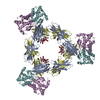
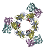
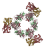
| タイトル | Structural analysis of a trimeric assembly of the mitochondrial dynamin-like GTPase Mgm1 |
|---|
 要素 要素 | Dynamin-like GTPase MGM1, mitochondrial |
|---|
 キーワード キーワード | HYDROLASE / Mitochondria / Fusion / Mgm1 |
|---|
| 機能・相同性 |  機能・相同性情報 機能・相同性情報
mitochondrial inner boundary membrane / Regulation of Apoptosis / mitochondrial outer membrane fusion / membrane bending / mitochondrion inheritance / mitochondrial inner membrane fusion / membrane tubulation / membrane bending activity / GTPase-dependent fusogenic activity / heme transport ...mitochondrial inner boundary membrane / Regulation of Apoptosis / mitochondrial outer membrane fusion / membrane bending / mitochondrion inheritance / mitochondrial inner membrane fusion / membrane tubulation / membrane bending activity / GTPase-dependent fusogenic activity / heme transport / dynamin GTPase / cristae formation / phosphatidic acid binding / : / cardiolipin binding / phosphatidylinositol-3,5-bisphosphate binding / mitochondrial membrane organization / mitochondrial fusion / phosphatidylserine binding / mitochondrial crista / mitochondrion organization / receptor internalization / mitochondrial intermembrane space / microtubule binding / mitochondrial outer membrane / microtubule / mitochondrial inner membrane / GTPase activity / GTP binding / mitochondrion / metal ion binding / plasma membrane / cytoplasm類似検索 - 分子機能 : / Dynamin-like GTPase MGM1-like, lipid interacting stalk / Dynamin, GTPase region, conserved site / Dynamin-type guanine nucleotide-binding (G) domain signature. / GTPase effector domain / GED domain profile. / Dynamin, GTPase domain / Dynamin, GTPase / Dynamin / Dynamin-type guanine nucleotide-binding (G) domain ...: / Dynamin-like GTPase MGM1-like, lipid interacting stalk / Dynamin, GTPase region, conserved site / Dynamin-type guanine nucleotide-binding (G) domain signature. / GTPase effector domain / GED domain profile. / Dynamin, GTPase domain / Dynamin, GTPase / Dynamin / Dynamin-type guanine nucleotide-binding (G) domain / Dynamin-type guanine nucleotide-binding (G) domain profile. / Dynamin, N-terminal / Dynamin family / P-loop containing nucleotide triphosphate hydrolases / Rossmann fold / P-loop containing nucleoside triphosphate hydrolase / 3-Layer(aba) Sandwich / Alpha Beta類似検索 - ドメイン・相同性 GUANOSINE-5'-DIPHOSPHATE / IODIDE ION / Dynamin-like GTPase MGM1, mitochondrial類似検索 - 構成要素 |
|---|
| 生物種 |   Saccharomyces cerevisiae S288c (パン酵母) Saccharomyces cerevisiae S288c (パン酵母) |
|---|
| 手法 |  X線回折 / シンクロトロン / 単波長異常分散 / 解像度: 3.2 Å X線回折 / シンクロトロン / 単波長異常分散 / 解像度: 3.2 Å |
|---|
 データ登録者 データ登録者 | Yan, L. / Li, L. |
|---|
| 資金援助 |  中国, 2件 中国, 2件 | 組織 | 認可番号 | 国 |
|---|
| Ministry of Science and Technology (MoST, China) | 2017YFC0840302 | 中国 | | National Natural Science Foundation of China (NSFC) | 31700659 | 中国 |
|
|---|
 引用 引用 | ジャーナル: Proc.Natl.Acad.Sci.USA / 年: 2020
タイトル: Structural analysis of a trimeric assembly of the mitochondrial dynamin-like GTPase Mgm1.
著者: Yan, L. / Qi, Y. / Ricketson, D. / Li, L. / Subramanian, K. / Zhao, J. / Yu, C. / Wu, L. / Sarsam, R. / Wong, M. / Lou, Z. / Rao, Z. / Nunnari, J. / Hu, J. |
|---|
| 履歴 | | 登録 | 2019年4月8日 | 登録サイト: PDBJ / 処理サイト: PDBJ |
|---|
| 改定 1.0 | 2020年2月19日 | Provider: repository / タイプ: Initial release |
|---|
| 改定 1.1 | 2020年2月26日 | Group: Database references / カテゴリ: citation / Item: _citation.pdbx_database_id_PubMed / _citation.title |
|---|
| 改定 1.2 | 2020年3月11日 | Group: Database references / カテゴリ: citation
Item: _citation.journal_volume / _citation.page_first / _citation.page_last |
|---|
| 改定 1.3 | 2024年11月20日 | Group: Data collection / Database references / Structure summary
カテゴリ: chem_comp_atom / chem_comp_bond ...chem_comp_atom / chem_comp_bond / database_2 / pdbx_entry_details / pdbx_modification_feature
Item: _database_2.pdbx_DOI / _database_2.pdbx_database_accession / _pdbx_entry_details.has_protein_modification |
|---|
|
|---|
 ムービー
ムービー コントローラー


 データを開く
 基本情報
基本情報 構造の表示
構造の表示 ダウンロードとリンク
 その他のダウンロード
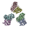

 PDBj
PDBj 集合体
集合体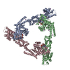


 タイプ: RNA linking / 分子量: 443.201 Da / 分子数: 3 / 由来タイプ: 合成 / 式: C10H15N5O11P2 / タイプ: SUBJECT OF INVESTIGATION / コメント: GDP, エネルギー貯蔵分子*YM
 分子量: 126.904 Da / 分子数: 25 / 由来タイプ: 合成 / 式: I
 試料調製
試料調製 解析
解析