- PDB-6jle: Crystal structure of MORN4/Myo3a complex -
+
Open data
ID or keywords:
Loading...
-
Basic information
Entry
Database: PDB / ID: 6jle
Title
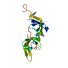
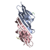
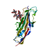
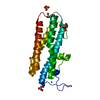
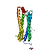
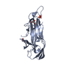
Crystal structure of MORN4/Myo3a complex
Components
MORN repeat-containing protein 4
Myosin-IIIa
Keywords
MOTOR PROTEIN / PROTEIN BINDING / PROTEIN TRANSPORT
Function / homology
Function and homology information
regulation of actin filament length / plus-end directed microfilament motor activity / cochlea morphogenesis / stereocilium tip / filopodium tip / myosin complex / positive regulation of filopodium assembly / Sensory processing of sound by outer hair cells of the cochlea / Sensory processing of sound by inner hair cells of the cochlea / microfilament motor activity ...regulation of actin filament length / plus-end directed microfilament motor activity / cochlea morphogenesis / stereocilium tip / filopodium tip / myosin complex / positive regulation of filopodium assembly / Sensory processing of sound by outer hair cells of the cochlea / Sensory processing of sound by inner hair cells of the cochlea / microfilament motor activity / filamentous actin / response to axon injury / cell projection / photoreceptor inner segment / visual perception / filopodium / sensory perception of sound / ADP binding / protein autophosphorylation / actin binding / protein kinase activity / calmodulin binding / non-specific serine/threonine protein kinase / protein serine kinase activity / protein serine/threonine kinase activity / ATP hydrolysis activity / ATP binding / cytoplasm Similarity search - Function
: / Class III myosin, motor domain / : / Possible plasma membrane-binding motif in junctophilins, PIP-5-kinases and protein kinases. / MORN motif / MORN repeat / IQ calmodulin-binding motif / Short calmodulin-binding motif containing conserved Ile and Gln residues. / IQ motif, EF-hand binding site / Myosin motor domain profile. ...: / Class III myosin, motor domain / : / Possible plasma membrane-binding motif in junctophilins, PIP-5-kinases and protein kinases. / MORN motif / MORN repeat / IQ calmodulin-binding motif / Short calmodulin-binding motif containing conserved Ile and Gln residues. / IQ motif, EF-hand binding site / Myosin motor domain profile. / Myosin head, motor domain / Myosin head (motor domain) / Myosin. Large ATPases. / IQ motif profile. / Kinesin motor domain superfamily / Protein kinase domain / Serine/Threonine protein kinases, catalytic domain / Protein kinase, ATP binding site / Protein kinases ATP-binding region signature. / Protein kinase domain profile. / Protein kinase domain / Protein kinase-like domain superfamily / P-loop containing nucleoside triphosphate hydrolase Similarity search - Domain/homology
Mass: 16226.919 Da / Num. of mol.: 1 Source method: isolated from a genetically manipulated source Source: (gene. exp.) Mus musculus (house mouse) / Gene: Morn4 / Production host: Escherichia coli (E. coli) / References: UniProt: Q6PGF2
#2: Protein/peptide
Myosin-IIIa
Mass: 5644.433 Da / Num. of mol.: 1 Source method: isolated from a genetically manipulated source Source: (gene. exp.) Homo sapiens (human) / Gene: MYO3A / Production host: Escherichia coli (E. coli) References: UniProt: Q8NEV4, non-specific serine/threonine protein kinase
In the structure databanks used in Yorodumi, some data are registered as the other names, "COVID-19 virus" and "2019-nCoV". Here are the details of the virus and the list of structure data.
Jan 31, 2019. EMDB accession codes are about to change! (news from PDBe EMDB page)
EMDB accession codes are about to change! (news from PDBe EMDB page)
The allocation of 4 digits for EMDB accession codes will soon come to an end. Whilst these codes will remain in use, new EMDB accession codes will include an additional digit and will expand incrementally as the available range of codes is exhausted. The current 4-digit format prefixed with “EMD-” (i.e. EMD-XXXX) will advance to a 5-digit format (i.e. EMD-XXXXX), and so on. It is currently estimated that the 4-digit codes will be depleted around Spring 2019, at which point the 5-digit format will come into force.
The EM Navigator/Yorodumi systems omit the EMD- prefix.
Related info.:Q: What is EMD? / ID/Accession-code notation in Yorodumi/EM Navigator
Yorodumi is a browser for structure data from EMDB, PDB, SASBDB, etc.
This page is also the successor to EM Navigator detail page, and also detail information page/front-end page for Omokage search.
The word "yorodu" (or yorozu) is an old Japanese word meaning "ten thousand". "mi" (miru) is to see.
Related info.:EMDB / PDB / SASBDB / Comparison of 3 databanks / Yorodumi Search / Aug 31, 2016. New EM Navigator & Yorodumi / Yorodumi Papers / Jmol/JSmol / Function and homology information / Changes in new EM Navigator and Yorodumi
 Movie
Movie Controller


 Open data
 Basic information
 Components
 Keywords
 Function and homology information
 Homo sapiens (human)
 X-RAY DIFFRACTION /
  Authors
 China, 1items
  Citation
 Structure visualization
 Downloads & links
 Other downloads
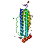


 PDBj
PDBj


 Assembly


 Mass: 92.094 Da / Num. of mol.: 3 / Source method: obtained synthetically / Formula: C3H8O3
 Mass: 192.124 Da / Num. of mol.: 2 / Source method: obtained synthetically / Formula: C6H8O7
 Mass: 18.015 Da / Num. of mol.: 133 / Source method: isolated from a natural source / Formula: H2O
 Sample preparation
 Processing