Protocol: SINGLE WAVELENGTH / Monochromatic (M) / Laue (L): M / Scattering type: x-ray
Radiation wavelength
Wavelength: 1.7 Å / Relative weight: 1
Reflection
Resolution: 1.06→30 Å / Num. obs: 17498 / % possible obs: 99.3 % / Redundancy: 21.2 % / CC1/2: 0.997 / Rmerge(I) obs: 0.036 / Net I/σ(I): 77.87
Reflection shell
Resolution: 1.06→1.1 Å / Redundancy: 19.1 % / Rmerge(I) obs: 0.213 / Mean I/σ(I) obs: 14.41 / Num. unique obs: 1685 / CC1/2: 0.993 / % possible all: 96.2
-
Processing
Software
Name
Version
Classification
PHENIX
1.13-2998
refinement
HKL-2000
datareduction
HKL-2000
datascaling
AutoSol
phasing
Refinement
Method to determine structure: SAD / Resolution: 1.06→21.25 Å / Cor.coef. Fo:Fc: 0.975 / Cor.coef. Fo:Fc free: 0.968 / SU B: 0.504 / SU ML: 0.012 / Cross valid method: THROUGHOUT / ESU R: 0.025 / ESU R Free: 0.026 Details: HYDROGENS HAVE BEEN ADDED IN THE RIDING POSITIONS U VALUES : REFINED INDIVIDUALLY
Rfactor
Num. reflection
% reflection
Selection details
Rfree
0.15798
895
5.1 %
RANDOM
Rwork
0.13577
-
-
-
obs
0.13695
16487
98.96 %
-
Solvent computation
Ion probe radii: 1 Å / Shrinkage radii: 1 Å / VDW probe radii: 1.7 Å
Displacement parameters
Biso mean: 17.174 Å2
Baniso -1
Baniso -2
Baniso -3
1-
-0.19 Å2
-0.09 Å2
-0 Å2
2-
-
-0.19 Å2
0 Å2
3-
-
-
0.61 Å2
Refinement step
Cycle: LAST / Resolution: 1.06→21.25 Å
Protein
Nucleic acid
Ligand
Solvent
Total
Num. atoms
314
0
10
43
367
Refine LS restraints
Refine-ID
Type
Dev ideal
Dev ideal target
Number
X-RAY DIFFRACTION
r_bond_refined_d
0.011
0.013
333
X-RAY DIFFRACTION
r_bond_other_d
0.001
0.017
277
X-RAY DIFFRACTION
r_angle_refined_deg
1.623
1.653
459
X-RAY DIFFRACTION
r_angle_other_deg
1.561
1.598
640
X-RAY DIFFRACTION
r_dihedral_angle_1_deg
7.555
5
39
X-RAY DIFFRACTION
r_dihedral_angle_2_deg
41.642
23.125
16
X-RAY DIFFRACTION
r_dihedral_angle_3_deg
8.25
15
42
X-RAY DIFFRACTION
r_dihedral_angle_4_deg
31.267
15
1
X-RAY DIFFRACTION
r_chiral_restr
0.081
0.2
41
X-RAY DIFFRACTION
r_gen_planes_refined
0.008
0.02
373
X-RAY DIFFRACTION
r_gen_planes_other
0.001
0.02
74
X-RAY DIFFRACTION
r_mcbond_it
1.032
1.572
159
X-RAY DIFFRACTION
r_mcbond_other
0.998
1.558
157
X-RAY DIFFRACTION
r_mcangle_it
1.462
2.362
197
X-RAY DIFFRACTION
r_mcangle_other
1.449
2.36
197
X-RAY DIFFRACTION
r_scbond_it
1.941
1.837
174
X-RAY DIFFRACTION
r_scbond_other
1.889
1.804
166
X-RAY DIFFRACTION
r_scangle_other
2.45
2.642
250
X-RAY DIFFRACTION
r_long_range_B_refined
2.811
19.645
364
X-RAY DIFFRACTION
r_long_range_B_other
2.478
18.629
351
X-RAY DIFFRACTION
r_rigid_bond_restr
1.722
3
610
LS refinement shell
Resolution: 1.062→1.089 Å / Total num. of bins used: 20
Rfactor
Num. reflection
% reflection
Rfree
0.145
47
-
Rwork
0.163
1156
-
obs
-
-
91.76 %
+
About Yorodumi
-
News
-
Feb 9, 2022. New format data for meta-information of EMDB entries
New format data for meta-information of EMDB entries
Version 3 of the EMDB header file is now the official format.
The previous official version 1.9 will be removed from the archive.
In the structure databanks used in Yorodumi, some data are registered as the other names, "COVID-19 virus" and "2019-nCoV". Here are the details of the virus and the list of structure data.
Jan 31, 2019. EMDB accession codes are about to change! (news from PDBe EMDB page)
EMDB accession codes are about to change! (news from PDBe EMDB page)
The allocation of 4 digits for EMDB accession codes will soon come to an end. Whilst these codes will remain in use, new EMDB accession codes will include an additional digit and will expand incrementally as the available range of codes is exhausted. The current 4-digit format prefixed with “EMD-” (i.e. EMD-XXXX) will advance to a 5-digit format (i.e. EMD-XXXXX), and so on. It is currently estimated that the 4-digit codes will be depleted around Spring 2019, at which point the 5-digit format will come into force.
The EM Navigator/Yorodumi systems omit the EMD- prefix.
Related info.:Q: What is EMD? / ID/Accession-code notation in Yorodumi/EM Navigator
Yorodumi is a browser for structure data from EMDB, PDB, SASBDB, etc.
This page is also the successor to EM Navigator detail page, and also detail information page/front-end page for Omokage search.
The word "yorodu" (or yorozu) is an old Japanese word meaning "ten thousand". "mi" (miru) is to see.
Related info.:EMDB / PDB / SASBDB / Comparison of 3 databanks / Yorodumi Search / Aug 31, 2016. New EM Navigator & Yorodumi / Yorodumi Papers / Jmol/JSmol / Function and homology information / Changes in new EM Navigator and Yorodumi
 Movie
Movie Controller


 Open data
 Basic information
 Components
 Keywords
 Function and homology information
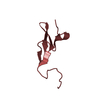
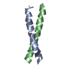
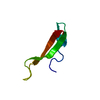
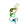
 Pholiota squarrosa (fungus)
 X-RAY DIFFRACTION /
  Authors
 Citation
 Structure visualization
 Downloads & links
 Other downloads
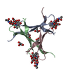


 PDBj
PDBj Assembly


 Mass: 96.063 Da / Num. of mol.: 2 / Source method: obtained synthetically / Formula: SO4
 Mass: 18.015 Da / Num. of mol.: 43 / Source method: isolated from a natural source / Formula: H2O
 Sample preparation
 / Beamline: TPS 05A / Wavelength: 1.7 Å
 Processing