 Movie
Movie Controller

[English] 日本語
 Yorodumi
- PDB-6jda: Crystal structure of N-acetyl mannosmaine kinase in complex with ... -
+ Open data

- Basic information
| Entry | Database: PDB / ID: 6jda | |||||||||
|---|---|---|---|---|---|---|---|---|---|---|
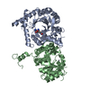
| Title | Crystal structure of N-acetyl mannosmaine kinase in complex with N-acetylmannosamine in Pasteurella multocida | |||||||||
 Components Components | N-acetylmannosamine kinase | |||||||||
 Keywords Keywords | SUGAR BINDING PROTEIN / Kinase / ManNAc binding protein / two domain protein | |||||||||
| Function / homology |  Function and homology information Function and homology informationN-acetylmannosamine metabolic process / N-acylmannosamine kinase / N-acylmannosamine kinase activity / N-acetylneuraminate catabolic process / zinc ion binding / ATP binding Similarity search - Function | |||||||||
| Biological species |  Pasteurella multocida (bacteria) Pasteurella multocida (bacteria) | |||||||||
| Method |  X-RAY DIFFRACTION / SYNCHROTRON / MOLECULAR REPLACEMENT / Resolution: 2.9 Å X-RAY DIFFRACTION / SYNCHROTRON / MOLECULAR REPLACEMENT / Resolution: 2.9 Å | |||||||||
 Authors Authors | Thanuja, G. / Ramaswamy, S. | |||||||||
| Funding support |  India, 2items India, 2items
| |||||||||
 Citation Citation | Journal: Acs Omega / Year: 2020 Title: Structure and Function of N‐Acetylmannosamine Kinases from Pathogenic Bacteria. Authors: Thanuja, G. / Ramaswamy, S. | |||||||||
| History |
|
- Structure visualization
| Structure viewer | Molecule: MolmilJmol/JSmol |
|---|
- Downloads & links
-Download
| PDBx/mmCIF format | 6jda.cif.gz | 69.7 KB | Display | PDBx/mmCIF format |
|---|---|---|---|---|
| PDB format | pdb6jda.ent.gz | 49.3 KB | Display | PDB format |
| PDBx/mmJSON format | 6jda.json.gz | Tree view | PDBx/mmJSON format | |
| Others |  Other downloads Other downloads |
-Validation report
| Arichive directory | https://data.pdbj.org/pub/pdb/validation_reports/jd/6jdaftp://data.pdbj.org/pub/pdb/validation_reports/jd/6jda | HTTPS FTP |
|---|
-Related structure data
| Related structure data |  6jdbC 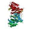 6jdcC  6jdhC  6jdoC  2aa4S S: Starting model for refinement C: citing same article ( |
|---|---|
| Similar structure data |


-Links
 PDBj
PDBj- Assembly
| Deposited unit | 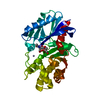
| ||||||||
|---|---|---|---|---|---|---|---|---|---|
| 1 | 
| ||||||||
| Unit cell |
|
-Components
| #1: Protein | Mass: 30689.037 Da / Num. of mol.: 1 Source method: isolated from a genetically manipulated source Details: ROK kinase / Source: (gene. exp.) Pasteurella multocida (bacteria) / Gene: nanK / Production host: References: UniProt: A0A2K0XYW4, UniProt: Q9CKB3*PLUS, N-acylmannosamine kinase |
|---|---|
| #2: Sugar | ChemComp-BM3 /   Type: D-saccharide, alpha linking / Mass: 221.208 Da / Num. of mol.: 1 Type: D-saccharide, alpha linking / Mass: 221.208 Da / Num. of mol.: 1Source method: isolated from a genetically manipulated source Formula: C8H15NO6 / Feature type: SUBJECT OF INVESTIGATION |
| #3: Chemical | ChemComp-ZN /   Mass: 65.409 Da / Num. of mol.: 1 / Source method: obtained synthetically / Formula: Zn / Feature type: SUBJECT OF INVESTIGATION Mass: 65.409 Da / Num. of mol.: 1 / Source method: obtained synthetically / Formula: Zn / Feature type: SUBJECT OF INVESTIGATION |
| #4: Chemical | ChemComp-GOL /   Mass: 92.094 Da / Num. of mol.: 1 / Source method: obtained synthetically / Formula: C3H8O3 Mass: 92.094 Da / Num. of mol.: 1 / Source method: obtained synthetically / Formula: C3H8O3 |
| #5: Water | ChemComp-HOH /  Mass: 18.015 Da / Num. of mol.: 7 / Source method: isolated from a natural source / Formula: H2O Mass: 18.015 Da / Num. of mol.: 7 / Source method: isolated from a natural source / Formula: H2O |
| Has ligand of interest | Y |
-Experimental details
-Experiment
| Experiment | Method: X-RAY DIFFRACTION / Number of used crystals: 1 |
|---|
- Sample preparation
| Crystal | Density Matthews: 3.53 Å3/Da / Density % sol: 65.2 % |
|---|---|
| Crystal grow | Temperature: 291 K / Method: vapor diffusion, hanging drop / pH: 7.5 Details: 0.2M lithium citrate tribasic tetrahydrate, 20%(w/v) PEG 3350 |
-Data collection
| Diffraction | Mean temperature: 100 K / Serial crystal experiment: N |
|---|---|
| Diffraction source | Source: SYNCHROTRON / Site: SOLEIL  / Beamline: PROXIMA 1 / Wavelength: 0.97857 Å / Beamline: PROXIMA 1 / Wavelength: 0.97857 Å |
| Detector | Type: DECTRIS PILATUS 6M / Detector: PIXEL / Date: Jul 1, 2016 |
| Radiation | Protocol: SINGLE WAVELENGTH / Monochromatic (M) / Laue (L): M / Scattering type: x-ray |
| Radiation wavelength | Wavelength: 0.97857 Å / Relative weight: 1 |
| Reflection | Resolution: 2.9→48.06 Å / Num. obs: 9983 / % possible obs: 99.5 % / Redundancy: 6.2 % / CC1/2: 0.995 / Rmerge(I) obs: 0.085 / Rpim(I) all: 0.038 / Rrim(I) all: 0.094 / Net I/σ(I): 13.4 / Num. measured all: 62338 / Scaling rejects: 28 |
| Reflection shell | Resolution: 2.9→3.08 Å / Redundancy: 6.3 % / Rmerge(I) obs: 0.447 / Num. measured all: 9925 / Num. unique obs: 1573 / CC1/2: 0.881 / Rpim(I) all: 0.196 / Rrim(I) all: 0.489 / Net I/σ(I) obs: 3.7 / % possible all: 99.8 |
- Processing
| Software |
| ||||||||||||||||||||||||||||
|---|---|---|---|---|---|---|---|---|---|---|---|---|---|---|---|---|---|---|---|---|---|---|---|---|---|---|---|---|---|
| Refinement | Method to determine structure: MOLECULAR REPLACEMENT Starting model: 2aa4 Resolution: 2.9→44.559 Å / SU ML: 0.37 / Cross valid method: THROUGHOUT / σ(F): 1.37 / Phase error: 27.85 / Stereochemistry target values: ML
| ||||||||||||||||||||||||||||
| Solvent computation | Shrinkage radii: 0.9 Å / VDW probe radii: 1.11 Å / Solvent model: FLAT BULK SOLVENT MODEL | ||||||||||||||||||||||||||||
| Displacement parameters | Biso max: 137.53 Å2 / Biso mean: 63.5832 Å2 / Biso min: 29.41 Å2 | ||||||||||||||||||||||||||||
| Refinement step | Cycle: final / Resolution: 2.9→44.559 Å
| ||||||||||||||||||||||||||||
| LS refinement shell | Refine-ID: X-RAY DIFFRACTION / Rfactor Rfree error: 0 / Total num. of bins used: 3
|
