 Movie
Movie Controller

[English] 日本語
 Yorodumi
- PDB-6jc9: Crystal structure of aminotransferase CrmG from Actinoalloteichus... -
+ Open data

- Basic information
| Entry | Database: PDB / ID: 6jc9 | ||||||
|---|---|---|---|---|---|---|---|
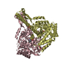
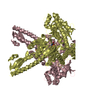
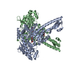
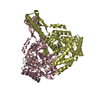
| Title | Crystal structure of aminotransferase CrmG from Actinoalloteichus sp. WH1-2216-6 in complex with amino donor L-Gln | ||||||
 Components Components | CrmG | ||||||
 Keywords Keywords | TRANSFERASE / aminotransferase / CrmG / complex | ||||||
| Function / homology |  Function and homology information Function and homology informationtransaminase activity / pyridoxal phosphate binding / identical protein binding Similarity search - Function | ||||||
| Biological species |  Actinoalloteichus sp. WH1-2216-6 (bacteria) Actinoalloteichus sp. WH1-2216-6 (bacteria) | ||||||
| Method |  X-RAY DIFFRACTION / SYNCHROTRON / MOLECULAR REPLACEMENT / molecular replacement / Resolution: 2.35 Å X-RAY DIFFRACTION / SYNCHROTRON / MOLECULAR REPLACEMENT / molecular replacement / Resolution: 2.35 Å | ||||||
 Authors Authors | Xu, J. / Liu, J. | ||||||
| Funding support |  China, 1items China, 1items
| ||||||
 Citation Citation | Journal: Commun Biol / Year: 2020 Title: Structural studies reveal flexible roof of active site responsible for omega-transaminase CrmG overcoming by-product inhibition. Authors: Xu, J. / Tang, X. / Zhu, Y. / Yu, Z. / Su, K. / Zhang, Y. / Dong, Y. / Zhu, W. / Zhang, C. / Wu, R. / Liu, J. | ||||||
| History |
|
- Structure visualization
| Structure viewer | Molecule: MolmilJmol/JSmol |
|---|
- Downloads & links
-Download
| PDBx/mmCIF format | 6jc9.cif.gz | 399.2 KB | Display | PDBx/mmCIF format |
|---|---|---|---|---|
| PDB format | pdb6jc9.ent.gz | 325.9 KB | Display | PDB format |
| PDBx/mmJSON format | 6jc9.json.gz | Tree view | PDBx/mmJSON format | |
| Others |  Other downloads Other downloads |
-Validation report
| Arichive directory | https://data.pdbj.org/pub/pdb/validation_reports/jc/6jc9ftp://data.pdbj.org/pub/pdb/validation_reports/jc/6jc9 | HTTPS FTP |
|---|
-Related structure data
| Related structure data |  6jc7C  6jc8C  6jcaC  6jcbC  5ddsS S: Starting model for refinement C: citing same article ( |
|---|---|
| Similar structure data |


-Links
 PDBj
PDBj

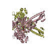
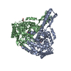
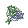
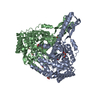
- Assembly
| Deposited unit | 
| ||||||||||||||||||||||||||||||||||||||||||||||||||||||||||||||||||||||||||||||||||||||||||||||||||
|---|---|---|---|---|---|---|---|---|---|---|---|---|---|---|---|---|---|---|---|---|---|---|---|---|---|---|---|---|---|---|---|---|---|---|---|---|---|---|---|---|---|---|---|---|---|---|---|---|---|---|---|---|---|---|---|---|---|---|---|---|---|---|---|---|---|---|---|---|---|---|---|---|---|---|---|---|---|---|---|---|---|---|---|---|---|---|---|---|---|---|---|---|---|---|---|---|---|---|---|
| 1 | 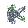
| ||||||||||||||||||||||||||||||||||||||||||||||||||||||||||||||||||||||||||||||||||||||||||||||||||
| 2 | 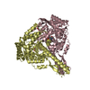
| ||||||||||||||||||||||||||||||||||||||||||||||||||||||||||||||||||||||||||||||||||||||||||||||||||
| Unit cell |
| ||||||||||||||||||||||||||||||||||||||||||||||||||||||||||||||||||||||||||||||||||||||||||||||||||
| Noncrystallographic symmetry (NCS) | NCS domain:
NCS domain segments: Component-ID: _ / Beg auth comp-ID: GLY / Beg label comp-ID: GLY / End auth comp-ID: LEU / End label comp-ID: LEU / Refine code: _ / Auth seq-ID: 6 - 522 / Label seq-ID: 6 - 522
NCS ensembles :
|
-Components
-Protein , 1 types, 4 molecules ABCD
| #1: Protein | Mass: 57219.520 Da / Num. of mol.: 4 Source method: isolated from a genetically manipulated source Source: (gene. exp.) Actinoalloteichus sp. WH1-2216-6 (bacteria)Production host: |
|---|
-Non-polymers , 5 types, 420 molecules 


| #2: Chemical | ChemComp-PLP /  Mass: 247.142 Da / Num. of mol.: 4 / Source method: obtained synthetically / Formula: C8H10NO6P Mass: 247.142 Da / Num. of mol.: 4 / Source method: obtained synthetically / Formula: C8H10NO6P#3: Chemical | ChemComp-ACY / |  Mass: 60.052 Da / Num. of mol.: 1 / Source method: obtained synthetically / Formula: C2H4O2 Mass: 60.052 Da / Num. of mol.: 1 / Source method: obtained synthetically / Formula: C2H4O2#4: Chemical | ChemComp-GOL / |  Mass: 92.094 Da / Num. of mol.: 1 / Source method: obtained synthetically / Formula: C3H8O3 Mass: 92.094 Da / Num. of mol.: 1 / Source method: obtained synthetically / Formula: C3H8O3#5: Chemical | ChemComp-GLN / |  Type: L-peptide linking / Mass: 146.144 Da / Num. of mol.: 1 / Source method: obtained synthetically / Formula: C5H10N2O3 Type: L-peptide linking / Mass: 146.144 Da / Num. of mol.: 1 / Source method: obtained synthetically / Formula: C5H10N2O3#6: Water | ChemComp-HOH / | Mass: 18.015 Da / Num. of mol.: 413 / Source method: isolated from a natural source / Formula: H2O |
|---|
-Experimental details
-Experiment
| Experiment | Method: X-RAY DIFFRACTION / Number of used crystals: 1 |
|---|
- Sample preparation
| Crystal | Density Matthews: 2.42 Å3/Da / Density % sol: 49.11 % / Mosaicity: 0.76 ° |
|---|---|
| Crystal grow | Temperature: 293 K / Method: vapor diffusion / pH: 8.5 Details: 0.2M Sodium acetate, 0.1M TRIS pH 8.5, 32% PEG 3350, 2% glycerol |
-Data collection
| Diffraction | Mean temperature: 100 K / Serial crystal experiment: N | ||||||||||||||||||||||||
|---|---|---|---|---|---|---|---|---|---|---|---|---|---|---|---|---|---|---|---|---|---|---|---|---|---|
| Diffraction source | Source: SYNCHROTRON / Site: SSRF / Beamline: BL17U1 / Wavelength: 0.9785 Å | ||||||||||||||||||||||||
| Detector | Type: ADSC QUANTUM 315r / Detector: CCD / Date: Oct 24, 2016 | ||||||||||||||||||||||||
| Radiation | Protocol: SINGLE WAVELENGTH / Monochromatic (M) / Laue (L): M / Scattering type: x-ray | ||||||||||||||||||||||||
| Radiation wavelength | Wavelength: 0.9785 Å / Relative weight: 1 | ||||||||||||||||||||||||
| Reflection | Resolution: 2.35→50.59 Å / Num. obs: 81461 / % possible obs: 91.2 % / Redundancy: 3.3 % / CC1/2: 0.992 / Rmerge(I) obs: 0.095 / Rpim(I) all: 0.061 / Rrim(I) all: 0.113 / Net I/σ(I): 9 | ||||||||||||||||||||||||
| Reflection shell | Diffraction-ID: 1
|
-Phasing
| Phasing | Method: molecular replacement |
|---|
- Processing
| Software |
| ||||||||||||||||||||||||||||||||||||||||||||||||||||||||||||
|---|---|---|---|---|---|---|---|---|---|---|---|---|---|---|---|---|---|---|---|---|---|---|---|---|---|---|---|---|---|---|---|---|---|---|---|---|---|---|---|---|---|---|---|---|---|---|---|---|---|---|---|---|---|---|---|---|---|---|---|---|---|
| Refinement | Method to determine structure: MOLECULAR REPLACEMENT Starting model: 5DDS Resolution: 2.35→50.59 Å / Cor.coef. Fo:Fc: 0.953 / Cor.coef. Fo:Fc free: 0.935 / SU B: 7.009 / SU ML: 0.162 / SU R Cruickshank DPI: 0.4874 / Cross valid method: THROUGHOUT / σ(F): 0 / ESU R: 0.487 / ESU R Free: 0.234 Details: SF FILE CONTAINS FRIEDEL PAIRS UNDER I/F_MINUS AND I/F_PLUS COLUMNS. HYDROGENS HAVE BEEN ADDED IN THE RIDING POSITIONS U VALUES : REFINED INDIVIDUALLY
| ||||||||||||||||||||||||||||||||||||||||||||||||||||||||||||
| Solvent computation | Ion probe radii: 0.8 Å / Shrinkage radii: 0.8 Å / VDW probe radii: 1.2 Å | ||||||||||||||||||||||||||||||||||||||||||||||||||||||||||||
| Displacement parameters | Biso max: 117.08 Å2 / Biso mean: 33.385 Å2 / Biso min: 11.45 Å2
| ||||||||||||||||||||||||||||||||||||||||||||||||||||||||||||
| Refinement step | Cycle: final / Resolution: 2.35→50.59 Å
| ||||||||||||||||||||||||||||||||||||||||||||||||||||||||||||
| Refine LS restraints |
| ||||||||||||||||||||||||||||||||||||||||||||||||||||||||||||
| Refine LS restraints NCS | Refine-ID: X-RAY DIFFRACTION / Type: interatomic distance / Rms dev position: 0.06 Å / Weight position: 0.05
| ||||||||||||||||||||||||||||||||||||||||||||||||||||||||||||
| LS refinement shell | Resolution: 2.35→2.411 Å / Rfactor Rfree error: 0 / Total num. of bins used: 20
|
