 Movie
Movie Controller

[English] 日本語
 Yorodumi
- PDB-6j35: Crystal structure of ligand-free of PulA-G680L mutant from Klebsi... -
+ Open data

- Basic information
| Entry | Database: PDB / ID: 6j35 | ||||||
|---|---|---|---|---|---|---|---|
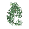
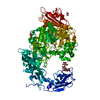
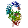
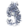
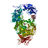
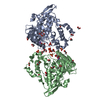
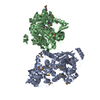
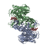
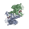
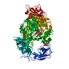
| Title | Crystal structure of ligand-free of PulA-G680L mutant from Klebsiella pneumoniae | ||||||
 Components Components | Pullulanase | ||||||
 Keywords Keywords | HYDROLASE / Pullulanase / Klebsiella pneumoniae / ligand-free / G680 | ||||||
| Function / homology |  Function and homology information Function and homology informationpullulanase / pullulanase activity / carbohydrate binding / carbohydrate metabolic process / metal ion binding Similarity search - Function | ||||||
| Biological species |  Klebsiella pneumoniae (bacteria) Klebsiella pneumoniae (bacteria) | ||||||
| Method |  X-RAY DIFFRACTION / SYNCHROTRON / MOLECULAR REPLACEMENT / Resolution: 1.839 Å X-RAY DIFFRACTION / SYNCHROTRON / MOLECULAR REPLACEMENT / Resolution: 1.839 Å | ||||||
 Authors Authors | Saka, N. / Iwamoto, H. / Takahashi, N. / Mizutani, K. / Mikami, B. | ||||||
| Funding support |  Japan, 1items Japan, 1items
| ||||||
 Citation Citation | Journal: Acta Crystallogr D Struct Biol / Year: 2019 Title: Relationship between the induced-fit loop and the activity of Klebsiella pneumoniae pullulanase. Authors: Saka, N. / Malle, D. / Iwamoto, H. / Takahashi, N. / Mizutani, K. / Mikami, B. | ||||||
| History |
|
- Structure visualization
| Structure viewer | Molecule: MolmilJmol/JSmol |
|---|
- Downloads & links
-Download
| PDBx/mmCIF format | 6j35.cif.gz | 453.9 KB | Display | PDBx/mmCIF format |
|---|---|---|---|---|
| PDB format | pdb6j35.ent.gz | 356.7 KB | Display | PDB format |
| PDBx/mmJSON format | 6j35.json.gz | Tree view | PDBx/mmJSON format | |
| Others |  Other downloads Other downloads |
-Validation report
| Arichive directory | https://data.pdbj.org/pub/pdb/validation_reports/j3/6j35ftp://data.pdbj.org/pub/pdb/validation_reports/j3/6j35 | HTTPS FTP |
|---|
-Related structure data
| Related structure data |  6j33C 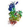 6j34C 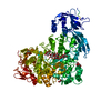 6j4hC 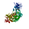 2fhfS S: Starting model for refinement C: citing same article ( |
|---|---|
| Similar structure data |
-Links
 PDBj
PDBj


- Assembly
| Deposited unit | 
| ||||||||
|---|---|---|---|---|---|---|---|---|---|
| 1 | 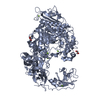
| ||||||||
| 2 | 
| ||||||||
| Unit cell |
|
-Components
| #1: Protein | Mass: 114518.805 Da / Num. of mol.: 2 / Mutation: G680L Source method: isolated from a genetically manipulated source Source: (gene. exp.) Klebsiella pneumoniae (bacteria) / Gene: pulA / Production host: #2: Chemical | ChemComp-MG /   Mass: 24.305 Da / Num. of mol.: 9 / Source method: obtained synthetically / Formula: Mg Mass: 24.305 Da / Num. of mol.: 9 / Source method: obtained synthetically / Formula: Mg#3: Chemical |   Mass: 92.094 Da / Num. of mol.: 2 / Source method: obtained synthetically / Formula: C3H8O3 Mass: 92.094 Da / Num. of mol.: 2 / Source method: obtained synthetically / Formula: C3H8O3#4: Chemical | ChemComp-CA / |   Mass: 40.078 Da / Num. of mol.: 1 / Source method: obtained synthetically / Formula: Ca Mass: 40.078 Da / Num. of mol.: 1 / Source method: obtained synthetically / Formula: Ca#5: Water | ChemComp-HOH / |  Mass: 18.015 Da / Num. of mol.: 1552 / Source method: isolated from a natural source / Formula: H2O Mass: 18.015 Da / Num. of mol.: 1552 / Source method: isolated from a natural source / Formula: H2OHas protein modification | Y | Sequence details | N229S was due to spontaneous mutation. | |
|---|
-Experimental details
-Experiment
| Experiment | Method: X-RAY DIFFRACTION / Number of used crystals: 1 |
|---|
- Sample preparation
| Crystal | Density Matthews: 2.38 Å3/Da / Density % sol: 48.32 % |
|---|---|
| Crystal grow | Temperature: 293 K / Method: vapor diffusion, hanging drop Details: 0.2M sodium acetate buffer (pH 6.0), 0.2M magnesium acetate, 20%(w/v) PEG 6000 |
-Data collection
| Diffraction | Mean temperature: 100 K / Serial crystal experiment: N |
|---|---|
| Diffraction source | Source: SYNCHROTRON / Site: SPring-8 / Beamline: BL44XU / Wavelength: 0.9 Å |
| Detector | Type: RAYONIX MX300HE / Detector: CCD / Date: Apr 16, 2017 |
| Radiation | Protocol: SINGLE WAVELENGTH / Monochromatic (M) / Laue (L): M / Scattering type: x-ray |
| Radiation wavelength | Wavelength: 0.9 Å / Relative weight: 1 |
| Reflection | Resolution: 1.839→50 Å / Num. obs: 181784 / % possible obs: 97.5 % / Redundancy: 2.7 % / Biso Wilson estimate: 16 Å2 / CC1/2: 0.997 / Rmerge(I) obs: 0.047 / Rpim(I) all: 0.037 / Rrim(I) all: 0.06 / Net I/av σ(I): 19.7 / Net I/σ(I): 19.7 |
| Reflection shell | Resolution: 1.84→1.9 Å / Redundancy: 2.7 % / Rmerge(I) obs: 0.136 / Mean I/σ(I) obs: 8.8 / Num. unique obs: 17267 / CC1/2: 0.968 / Rpim(I) all: 0.107 / Rrim(I) all: 0.175 / % possible all: 93.3 |
- Processing
| Software |
| |||||||||||||||||||||||||||||||||||||||||||||||||||||||||||||||||||||||||||||||||||||||||||||||||||||||||||||||||||||||||||||||||||||||||||||||||||||||||||||||||||||||||||||||||||||||||||||||||||||||||||||||||||||||||
|---|---|---|---|---|---|---|---|---|---|---|---|---|---|---|---|---|---|---|---|---|---|---|---|---|---|---|---|---|---|---|---|---|---|---|---|---|---|---|---|---|---|---|---|---|---|---|---|---|---|---|---|---|---|---|---|---|---|---|---|---|---|---|---|---|---|---|---|---|---|---|---|---|---|---|---|---|---|---|---|---|---|---|---|---|---|---|---|---|---|---|---|---|---|---|---|---|---|---|---|---|---|---|---|---|---|---|---|---|---|---|---|---|---|---|---|---|---|---|---|---|---|---|---|---|---|---|---|---|---|---|---|---|---|---|---|---|---|---|---|---|---|---|---|---|---|---|---|---|---|---|---|---|---|---|---|---|---|---|---|---|---|---|---|---|---|---|---|---|---|---|---|---|---|---|---|---|---|---|---|---|---|---|---|---|---|---|---|---|---|---|---|---|---|---|---|---|---|---|---|---|---|---|---|---|---|---|---|---|---|---|---|---|---|---|---|---|---|---|
| Refinement | Method to determine structure: MOLECULAR REPLACEMENT Starting model: 2FHF Resolution: 1.839→38.721 Å / SU ML: 0.15 / Cross valid method: FREE R-VALUE / σ(F): 1.97 / Phase error: 21.97 / Stereochemistry target values: ML
| |||||||||||||||||||||||||||||||||||||||||||||||||||||||||||||||||||||||||||||||||||||||||||||||||||||||||||||||||||||||||||||||||||||||||||||||||||||||||||||||||||||||||||||||||||||||||||||||||||||||||||||||||||||||||
| Solvent computation | Shrinkage radii: 0.9 Å / VDW probe radii: 1.11 Å / Solvent model: FLAT BULK SOLVENT MODEL | |||||||||||||||||||||||||||||||||||||||||||||||||||||||||||||||||||||||||||||||||||||||||||||||||||||||||||||||||||||||||||||||||||||||||||||||||||||||||||||||||||||||||||||||||||||||||||||||||||||||||||||||||||||||||
| Refinement step | Cycle: LAST / Resolution: 1.839→38.721 Å
| |||||||||||||||||||||||||||||||||||||||||||||||||||||||||||||||||||||||||||||||||||||||||||||||||||||||||||||||||||||||||||||||||||||||||||||||||||||||||||||||||||||||||||||||||||||||||||||||||||||||||||||||||||||||||
| Refine LS restraints |
| |||||||||||||||||||||||||||||||||||||||||||||||||||||||||||||||||||||||||||||||||||||||||||||||||||||||||||||||||||||||||||||||||||||||||||||||||||||||||||||||||||||||||||||||||||||||||||||||||||||||||||||||||||||||||
| LS refinement shell |
|
