 Movie
Movie Controller

[English] 日本語
 Yorodumi
- PDB-6iqh: X-ray crystal structure of covalent-bonded complex of Fc and peptide -
+ Open data

- Basic information
| Entry | Database: PDB / ID: 6iqh | ||||||||||||
|---|---|---|---|---|---|---|---|---|---|---|---|---|---|
| Title | X-ray crystal structure of covalent-bonded complex of Fc and peptide | ||||||||||||
 Components Components |
| ||||||||||||
 Keywords Keywords | IMMUNE SYSTEM / Fc / Complex | ||||||||||||
| Function / homology |  Function and homology information Function and homology informationimmunoglobulin complex / adaptive immune response / extracellular region / plasma membrane Similarity search - Function | ||||||||||||
| Biological species |  Homo sapiens (human) Homo sapiens (human)synthetic construct (others) | ||||||||||||
| Method |  X-RAY DIFFRACTION / SYNCHROTRON / FOURIER SYNTHESIS / Resolution: 2.999 Å X-RAY DIFFRACTION / SYNCHROTRON / FOURIER SYNTHESIS / Resolution: 2.999 Å | ||||||||||||
 Authors Authors | Adachi, M. / Ito, Y. | ||||||||||||
 Citation Citation | Journal: Bioconjug. Chem. / Year: 2019 Title: Site-Specific Chemical Conjugation of Antibodies by Using Affinity Peptide for the Development of Therapeutic Antibody Format. Authors: Kishimoto, S. / Nakashimada, Y. / Yokota, R. / Hatanaka, T. / Adachi, M. / Ito, Y. | ||||||||||||
| History |
|
- Structure visualization
| Structure viewer | Molecule: MolmilJmol/JSmol |
|---|
- Downloads & links
-Download
| PDBx/mmCIF format | 6iqh.cif.gz | 109.6 KB | Display | PDBx/mmCIF format |
|---|---|---|---|---|
| PDB format | pdb6iqh.ent.gz | Display | PDB format | |
| PDBx/mmJSON format | 6iqh.json.gz | Tree view | PDBx/mmJSON format | |
| Others |  Other downloads Other downloads |
-Validation report
| Arichive directory | https://data.pdbj.org/pub/pdb/validation_reports/iq/6iqhftp://data.pdbj.org/pub/pdb/validation_reports/iq/6iqh | HTTPS FTP |
|---|
-Related structure data
| Related structure data | 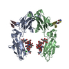 6iqgC 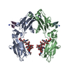 1dn2S S: Starting model for refinement C: citing same article ( |
|---|---|
| Similar structure data |


-Links
 PDBj
PDBj


- Assembly
| Deposited unit | 
| ||||||||
|---|---|---|---|---|---|---|---|---|---|
| 1 |
| ||||||||
| Unit cell |
|
-Components
| #1: Antibody | Mass: 23788.842 Da / Num. of mol.: 2 Source method: isolated from a genetically manipulated source Source: (gene. exp.) Homo sapiens (human) / Production host:   Cricetulus griseus (Chinese hamster) / References: UniProt: P0DOX5 Cricetulus griseus (Chinese hamster) / References: UniProt: P0DOX5#2: Protein/peptide | Mass: 2063.314 Da / Num. of mol.: 2 / Source method: obtained synthetically / Source: (synth.) synthetic construct (others) #3: Polysaccharide | Source method: isolated from a genetically manipulated source |
|---|
-Experimental details
-Experiment
| Experiment | Method: X-RAY DIFFRACTION / Number of used crystals: 1 |
|---|
- Sample preparation
| Crystal | Density Matthews: 3.18 Å3/Da / Density % sol: 61.33 % |
|---|---|
| Crystal grow | Temperature: 293 K / Method: vapor diffusion, hanging drop / pH: 10 / Details: 0.1 M CHES pH 10.0, 0.3M NaCl, 20% (w/v) PEG 8000 |
-Data collection
| Diffraction | Mean temperature: 100 K / Serial crystal experiment: N |
|---|---|
| Diffraction source | Source: SYNCHROTRON / Site: Photon Factory  / Beamline: BL-17A / Wavelength: 0.98 Å / Beamline: BL-17A / Wavelength: 0.98 Å |
| Detector | Type: DECTRIS PILATUS3 6M / Detector: PIXEL / Date: May 24, 2016 |
| Radiation | Protocol: SINGLE WAVELENGTH / Monochromatic (M) / Laue (L): M / Scattering type: x-ray |
| Radiation wavelength | Wavelength: 0.98 Å / Relative weight: 1 |
| Reflection | Resolution: 2.999→44.1 Å / Num. obs: 12253 / % possible obs: 98.4 % / Redundancy: 3.1 % / Rmerge(I) obs: 0.159 / Net I/σ(I): 8.4 |
| Reflection shell | Resolution: 3→3.11 Å / Redundancy: 2.6 % / Rmerge(I) obs: 0.417 / Mean I/σ(I) obs: 1.6 / Num. unique obs: 1158 / % possible all: 92.9 |
- Processing
| Software |
| |||||||||||||||||||||||||||||||||||
|---|---|---|---|---|---|---|---|---|---|---|---|---|---|---|---|---|---|---|---|---|---|---|---|---|---|---|---|---|---|---|---|---|---|---|---|---|
| Refinement | Method to determine structure: FOURIER SYNTHESIS Starting model: 1DN2 Resolution: 2.999→44.1 Å / SU ML: 0.47 / Cross valid method: FREE R-VALUE / σ(F): 1.36 / Phase error: 34.78
| |||||||||||||||||||||||||||||||||||
| Solvent computation | Shrinkage radii: 0.9 Å / VDW probe radii: 1.11 Å | |||||||||||||||||||||||||||||||||||
| Refinement step | Cycle: LAST / Resolution: 2.999→44.1 Å
| |||||||||||||||||||||||||||||||||||
| Refine LS restraints |
| |||||||||||||||||||||||||||||||||||
| LS refinement shell |
|
