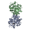
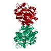
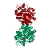
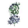
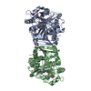
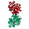
Evidence: gel filtration, The apoprotein crystallised as two hsEH CTD molecules in the asymmetric unit forming a homodimer. However, gel filtration analysis showed isolated monodispersed and ...Evidence: gel filtration, The apoprotein crystallised as two hsEH CTD molecules in the asymmetric unit forming a homodimer. However, gel filtration analysis showed isolated monodispersed and monomeric protein in solution.
Type
Name
Symmetry operation
Number
identity operation
1_555
x,y,z
1
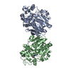
Buried area
1450 Å2
ΔGint
-1 kcal/mol
Surface area
25070 Å2
Method
PISA
Unit cell
Length a, b, c (Å)
88.224, 80.139, 104.696
Angle α, β, γ (deg.)
90.00, 95.39, 90.00
Int Tables number
5
Space group name H-M
I121
-
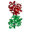
Components
#1: Protein
Bifunctionalepoxidehydrolase2
Mass: 39545.250 Da / Num. of mol.: 2 Source method: isolated from a genetically manipulated source Source: (gene. exp.) Homo sapiens (human) / Gene: EPHX2 / Production host: Escherichia coli (E. coli) References: UniProt: P34913, soluble epoxide hydrolase, lipid-phosphate phosphatase
Resolution: 2.6→70.51 Å / Cor.coef. Fo:Fc: 0.91 / Cor.coef. Fo:Fc free: 0.886 / SU B: 20.645 / SU ML: 0.393 / Cross valid method: THROUGHOUT / ESU R: 1.475 / ESU R Free: 0.358 / Details: HYDROGENS HAVE BEEN ADDED IN THE RIDING POSITIONS
Rfactor
Num. reflection
% reflection
Selection details
Rfree
0.27704
1101
4.9 %
RANDOM
Rwork
0.24599
-
-
-
obs
0.24755
21388
99.9 %
-
Solvent computation
Ion probe radii: 0.8 Å / Shrinkage radii: 0.8 Å / VDW probe radii: 1.2 Å
 Movie
Movie Controller

 Yorodumi
 Open data
 Basic information
 Components
 Keywords
 Function and homology information
 Homo sapiens (human)
 X-RAY DIFFRACTION /
  Authors
 United Kingdom, 1items
  Citation
 Structure visualization
 Downloads & links
 Other downloads
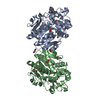


 PDBj
PDBj


 Assembly
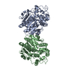

 Mass: 18.015 Da / Num. of mol.: 13 / Source method: isolated from a natural source / Formula: H2O
 Sample preparation
 Processing