 Movie
Movie Controller

[English] 日本語
 Yorodumi
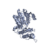
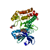
- PDB-6htj: Crystal structure of the translation recovery factor Trf from Sul... -
+ Open data

- Basic information
| Entry | Database: PDB / ID: 6htj | ||||||
|---|---|---|---|---|---|---|---|
| Title | Crystal structure of the translation recovery factor Trf from Sulfolobus solfataricus | ||||||
 Components Components | Nucleic-acid-binding protein containing a Zn-ribbon | ||||||
 Keywords Keywords | TRANSLATION / translation initiation / ribosome / Sulfolobus solfataricus / translation recovery factor Trf | ||||||
| Function / homology | : / Domain of unknown function DUF35, rubredoxin-like zinc ribbon domain, N-terminal / ChsH2, rubredoxin-like zinc ribbon domain / Nucleic acid-binding, OB-fold / metal ion binding / Zn-ribbon domain-containing OB-fold protein / DUF35 domain-containing protein Function and homology information Function and homology information | ||||||
| Biological species |   Sulfolobus solfataricus (archaea) Sulfolobus solfataricus (archaea) | ||||||
| Method |  X-RAY DIFFRACTION / SYNCHROTRON / SAD / Resolution: 1.65 Å X-RAY DIFFRACTION / SYNCHROTRON / SAD / Resolution: 1.65 Å | ||||||
 Authors Authors | Woehnert, J. / Pogoryelov, D. / Kaiser, M. | ||||||
 Citation Citation | Journal: Febs Open Bio / Year: 2019 Title: Crystal structure of the translation recovery factor Trf from Sulfolobus solfataricus. Authors: Kaiser, M. / Wurm, J.P. / Martens, B. / Blasi, U. / Pogoryelov, D. / Wohnert, J. | ||||||
| History |
|
- Structure visualization
| Structure viewer | Molecule: MolmilJmol/JSmol |
|---|
- Downloads & links
-Download
| PDBx/mmCIF format | 6htj.cif.gz | 68.1 KB | Display | PDBx/mmCIF format |
|---|---|---|---|---|
| PDB format | pdb6htj.ent.gz | 49.5 KB | Display | PDB format |
| PDBx/mmJSON format | 6htj.json.gz | Tree view | PDBx/mmJSON format | |
| Others |  Other downloads Other downloads |
-Validation report
| Summary document | 6htj_validation.pdf.gz | 426.3 KB | Display | wwPDB validaton report |
|---|---|---|---|---|
| Full document | 6htj_full_validation.pdf.gz | 426.6 KB | Display | |
| Data in XML | 6htj_validation.xml.gz | 13.7 KB | Display | |
| Data in CIF | 6htj_validation.cif.gz | 19.9 KB | Display | |
| Arichive directory | https://data.pdbj.org/pub/pdb/validation_reports/ht/6htjftp://data.pdbj.org/pub/pdb/validation_reports/ht/6htj | HTTPS FTP |
-Related structure data
| Similar structure data |
|---|


-Links
 PDBj
PDBj
- Assembly
| Deposited unit | 
| ||||||||
|---|---|---|---|---|---|---|---|---|---|
| 1 |
| ||||||||
| Unit cell |
|
-Components
| #1: Protein | Mass: 14159.465 Da / Num. of mol.: 2 Source method: isolated from a genetically manipulated source Source: (gene. exp.) Sulfolobus solfataricus (archaea) / Gene: SSOP1_2634, SULA_0310, SULB_0312, SULC_0310 / Production host:  #2: Chemical |   Mass: 65.409 Da / Num. of mol.: 2 / Source method: obtained synthetically / Formula: Zn Mass: 65.409 Da / Num. of mol.: 2 / Source method: obtained synthetically / Formula: Zn#3: Water | ChemComp-HOH / |  Mass: 18.015 Da / Num. of mol.: 253 / Source method: isolated from a natural source / Formula: H2O Mass: 18.015 Da / Num. of mol.: 253 / Source method: isolated from a natural source / Formula: H2O |
|---|
-Experimental details
-Experiment
| Experiment | Method: X-RAY DIFFRACTION / Number of used crystals: 1 |
|---|
- Sample preparation
| Crystal | Density Matthews: 2.11 Å3/Da / Density % sol: 41.68 % |
|---|---|
| Crystal grow | Temperature: 277.5 K / Method: microdialysis / pH: 6.5 Details: 50 mM Bis Tris pH 6.5, 50 mM NaCl, 5 mM beta-mercapthoethanol |
-Data collection
| Diffraction | Mean temperature: 100 K / Serial crystal experiment: N |
|---|---|
| Diffraction source | Source: SYNCHROTRON / Site: SLS  / Beamline: X06SA / Wavelength: 1.00004 Å / Beamline: X06SA / Wavelength: 1.00004 Å |
| Detector | Type: DECTRIS PILATUS 2M / Detector: PIXEL / Date: May 14, 2014 |
| Radiation | Protocol: SINGLE WAVELENGTH / Monochromatic (M) / Laue (L): M / Scattering type: x-ray |
| Radiation wavelength | Wavelength: 1.00004 Å / Relative weight: 1 |
| Reflection | Resolution: 1.65→38.304 Å / Num. obs: 29528 / % possible obs: 99.82 % / Redundancy: 13.5 % / Biso Wilson estimate: 17.8 Å2 / Rmerge(I) obs: 0.1447 / Rpim(I) all: 0.04051 / Rrim(I) all: 0.1504 / Net I/σ(I): 13.32 |
| Reflection shell | Resolution: 1.65→1.709 Å / Redundancy: 12.1 % / Rmerge(I) obs: 1.238 / Num. unique obs: 2817 / Rpim(I) all: 0.3628 / Rrim(I) all: 1.292 / % possible all: 98.26 |
-Phasing
| Phasing | Method: SAD |
|---|
- Processing
| Software |
| |||||||||||||||||||||||||||||||||||||||||||||||||||||||||||||||||||||||||||||||||||||||||||||||||||||||||||||||||||||||||||||||||||||||||||||||||||
|---|---|---|---|---|---|---|---|---|---|---|---|---|---|---|---|---|---|---|---|---|---|---|---|---|---|---|---|---|---|---|---|---|---|---|---|---|---|---|---|---|---|---|---|---|---|---|---|---|---|---|---|---|---|---|---|---|---|---|---|---|---|---|---|---|---|---|---|---|---|---|---|---|---|---|---|---|---|---|---|---|---|---|---|---|---|---|---|---|---|---|---|---|---|---|---|---|---|---|---|---|---|---|---|---|---|---|---|---|---|---|---|---|---|---|---|---|---|---|---|---|---|---|---|---|---|---|---|---|---|---|---|---|---|---|---|---|---|---|---|---|---|---|---|---|---|---|---|---|
| Refinement | Method to determine structure: SAD / Resolution: 1.65→38.304 Å / SU ML: 0.18 / Cross valid method: FREE R-VALUE / σ(F): 1.36 / Phase error: 20.06
| |||||||||||||||||||||||||||||||||||||||||||||||||||||||||||||||||||||||||||||||||||||||||||||||||||||||||||||||||||||||||||||||||||||||||||||||||||
| Solvent computation | Shrinkage radii: 0.9 Å / VDW probe radii: 1.11 Å | |||||||||||||||||||||||||||||||||||||||||||||||||||||||||||||||||||||||||||||||||||||||||||||||||||||||||||||||||||||||||||||||||||||||||||||||||||
| Displacement parameters | Biso max: 65.89 Å2 / Biso mean: 25.7392 Å2 / Biso min: 10.67 Å2 | |||||||||||||||||||||||||||||||||||||||||||||||||||||||||||||||||||||||||||||||||||||||||||||||||||||||||||||||||||||||||||||||||||||||||||||||||||
| Refinement step | Cycle: final / Resolution: 1.65→38.304 Å
| |||||||||||||||||||||||||||||||||||||||||||||||||||||||||||||||||||||||||||||||||||||||||||||||||||||||||||||||||||||||||||||||||||||||||||||||||||
| Refine LS restraints |
| |||||||||||||||||||||||||||||||||||||||||||||||||||||||||||||||||||||||||||||||||||||||||||||||||||||||||||||||||||||||||||||||||||||||||||||||||||
| LS refinement shell | Refine-ID: X-RAY DIFFRACTION / Rfactor Rfree error: 0 / Total num. of bins used: 20
|
