 Movie
Movie Controller


+ Open data

- Basic information
| Entry | Database: PDB / ID: 6hnc | ||||||
|---|---|---|---|---|---|---|---|
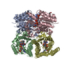
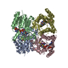
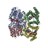
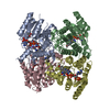
| Title | Trypanosoma brucei PTR1 in complex with cycloguanil | ||||||
 Components Components | Pteridine reductase | ||||||
 Keywords Keywords | OXIDOREDUCTASE / Trypanosoma brucei / pteridine reductase / PTR1 / TbPTR1 / cycloguanil | ||||||
| Function / homology |  Function and homology information Function and homology information | ||||||
| Biological species |  | ||||||
| Method |  X-RAY DIFFRACTION / SYNCHROTRON / MOLECULAR REPLACEMENT / Resolution: 1.5 Å X-RAY DIFFRACTION / SYNCHROTRON / MOLECULAR REPLACEMENT / Resolution: 1.5 Å | ||||||
 Authors Authors | Landi, G. / Pozzi, C. / Mangani, S. | ||||||
| Funding support | 1items
| ||||||
 Citation Citation | Journal: Acs Infect Dis. / Year: 2019 Title: Structural Insights into the Development of Cycloguanil Derivatives asTrypanosoma bruceiPteridine-Reductase-1 Inhibitors. Authors: Landi, G. / Linciano, P. / Borsari, C. / Bertolacini, C.P. / Moraes, C.B. / Cordeiro-da-Silva, A. / Gul, S. / Witt, G. / Kuzikov, M. / Costi, M.P. / Pozzi, C. / Mangani, S. | ||||||
| History |
|
- Structure visualization
| Structure viewer | Molecule: MolmilJmol/JSmol |
|---|
- Downloads & links
-Download
| PDBx/mmCIF format | 6hnc.cif.gz | 228.4 KB | Display | PDBx/mmCIF format |
|---|---|---|---|---|
| PDB format | pdb6hnc.ent.gz | 180.1 KB | Display | PDB format |
| PDBx/mmJSON format | 6hnc.json.gz | Tree view | PDBx/mmJSON format | |
| Others |  Other downloads Other downloads |
-Validation report
| Arichive directory | https://data.pdbj.org/pub/pdb/validation_reports/hn/6hncftp://data.pdbj.org/pub/pdb/validation_reports/hn/6hnc | HTTPS FTP |
|---|
-Related structure data
| Related structure data |  6hnrC  6howC 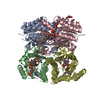 5jdcS S: Starting model for refinement C: citing same article ( |
|---|---|
| Similar structure data |


-Links
 PDBj
PDBj


- Assembly
| Deposited unit | 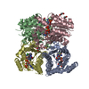
| ||||||||
|---|---|---|---|---|---|---|---|---|---|
| 1 |
| ||||||||
| Unit cell |
|
-Components
-Protein , 1 types, 4 molecules ABCD
| #1: Protein | Mass: 30669.791 Da / Num. of mol.: 4 Source method: isolated from a genetically manipulated source Source: (gene. exp.)  |
|---|
-Non-polymers , 6 types, 833 molecules 


| #2: Chemical | ChemComp-NAP /  Mass: 743.405 Da / Num. of mol.: 4 / Source method: obtained synthetically / Formula: C21H28N7O17P3 Mass: 743.405 Da / Num. of mol.: 4 / Source method: obtained synthetically / Formula: C21H28N7O17P3#3: Chemical |  Mass: 251.715 Da / Num. of mol.: 3 / Source method: obtained synthetically / Formula: C11H14ClN5 / Comment: inhibitor, antagonist*YM Mass: 251.715 Da / Num. of mol.: 3 / Source method: obtained synthetically / Formula: C11H14ClN5 / Comment: inhibitor, antagonist*YM#4: Chemical | ChemComp-ACT /  Mass: 59.044 Da / Num. of mol.: 4 / Source method: obtained synthetically / Formula: C2H3O2 Mass: 59.044 Da / Num. of mol.: 4 / Source method: obtained synthetically / Formula: C2H3O2#5: Chemical | ChemComp-GOL /  Mass: 92.094 Da / Num. of mol.: 5 / Source method: obtained synthetically / Formula: C3H8O3 Mass: 92.094 Da / Num. of mol.: 5 / Source method: obtained synthetically / Formula: C3H8O3#6: Chemical |  Mass: 78.133 Da / Num. of mol.: 2 / Source method: obtained synthetically / Formula: C2H6OS / Comment: DMSO, precipitant*YM Mass: 78.133 Da / Num. of mol.: 2 / Source method: obtained synthetically / Formula: C2H6OS / Comment: DMSO, precipitant*YM#7: Water | ChemComp-HOH / | Mass: 18.015 Da / Num. of mol.: 815 / Source method: isolated from a natural source / Formula: H2O |
|---|
-Experimental details
-Experiment
| Experiment | Method: X-RAY DIFFRACTION / Number of used crystals: 1 |
|---|
- Sample preparation
| Crystal | Density Matthews: 2.09 Å3/Da / Density % sol: 41.19 % |
|---|---|
| Crystal grow | Temperature: 294 K / Method: vapor diffusion, hanging drop / pH: 5 / Details: 2-2.5 M sodium acetate, 0.1 M sodium citrate, pH5 |
-Data collection
| Diffraction | Mean temperature: 100 K |
|---|---|
| Diffraction source | Source: SYNCHROTRON / Site: ESRF  / Beamline: ID30B / Wavelength: 0.96863 Å / Beamline: ID30B / Wavelength: 0.96863 Å |
| Detector | Type: DECTRIS PILATUS3 6M / Detector: PIXEL / Date: May 10, 2017 |
| Radiation | Protocol: SINGLE WAVELENGTH / Monochromatic (M) / Laue (L): M / Scattering type: x-ray |
| Radiation wavelength | Wavelength: 0.96863 Å / Relative weight: 1 |
| Reflection | Resolution: 1.5→74.76 Å / Num. obs: 152088 / % possible obs: 96.2 % / Observed criterion σ(I): 2 / Redundancy: 3.1 % / Biso Wilson estimate: 13.6 Å2 / CC1/2: 0.997 / Rmerge(I) obs: 0.059 / Rpim(I) all: 0.039 / Rrim(I) all: 0.072 / Net I/σ(I): 7.8 |
| Reflection shell | Resolution: 1.5→1.58 Å / Redundancy: 2.9 % / Rmerge(I) obs: 0.453 / Mean I/σ(I) obs: 2 / Num. unique obs: 21865 / CC1/2: 0.864 / Rpim(I) all: 0.312 / Rrim(I) all: 0.553 / % possible all: 94.9 |
- Processing
| Software |
| ||||||||||||||||||||||||||||||||||||||||||||||||||||||||||||||||||||||||||||||||||||||||||||||||||||||||||||||||||||||||||||||||||||||||||||||||||||||||||||||||||||||||||||||||||||||
|---|---|---|---|---|---|---|---|---|---|---|---|---|---|---|---|---|---|---|---|---|---|---|---|---|---|---|---|---|---|---|---|---|---|---|---|---|---|---|---|---|---|---|---|---|---|---|---|---|---|---|---|---|---|---|---|---|---|---|---|---|---|---|---|---|---|---|---|---|---|---|---|---|---|---|---|---|---|---|---|---|---|---|---|---|---|---|---|---|---|---|---|---|---|---|---|---|---|---|---|---|---|---|---|---|---|---|---|---|---|---|---|---|---|---|---|---|---|---|---|---|---|---|---|---|---|---|---|---|---|---|---|---|---|---|---|---|---|---|---|---|---|---|---|---|---|---|---|---|---|---|---|---|---|---|---|---|---|---|---|---|---|---|---|---|---|---|---|---|---|---|---|---|---|---|---|---|---|---|---|---|---|---|---|
| Refinement | Method to determine structure: MOLECULAR REPLACEMENT Starting model: 5JDC Resolution: 1.5→66.51 Å / Cor.coef. Fo:Fc: 0.968 / Cor.coef. Fo:Fc free: 0.956 / SU B: 1.776 / SU ML: 0.062 / SU R Cruickshank DPI: 0.076 / Cross valid method: THROUGHOUT / ESU R: 0.076 / ESU R Free: 0.079 / SU Rfree Cruickshank DPI: 0.079
| ||||||||||||||||||||||||||||||||||||||||||||||||||||||||||||||||||||||||||||||||||||||||||||||||||||||||||||||||||||||||||||||||||||||||||||||||||||||||||||||||||||||||||||||||||||||
| Solvent computation | Ion probe radii: 0.8 Å / Shrinkage radii: 0.8 Å / VDW probe radii: 1.2 Å | ||||||||||||||||||||||||||||||||||||||||||||||||||||||||||||||||||||||||||||||||||||||||||||||||||||||||||||||||||||||||||||||||||||||||||||||||||||||||||||||||||||||||||||||||||||||
| Displacement parameters | Biso mean: 19.309 Å2
| ||||||||||||||||||||||||||||||||||||||||||||||||||||||||||||||||||||||||||||||||||||||||||||||||||||||||||||||||||||||||||||||||||||||||||||||||||||||||||||||||||||||||||||||||||||||
| Refine analyze | Luzzati coordinate error obs: 0.174 Å | ||||||||||||||||||||||||||||||||||||||||||||||||||||||||||||||||||||||||||||||||||||||||||||||||||||||||||||||||||||||||||||||||||||||||||||||||||||||||||||||||||||||||||||||||||||||
| Refinement step | Cycle: 1 / Resolution: 1.5→66.51 Å
| ||||||||||||||||||||||||||||||||||||||||||||||||||||||||||||||||||||||||||||||||||||||||||||||||||||||||||||||||||||||||||||||||||||||||||||||||||||||||||||||||||||||||||||||||||||||
| Refine LS restraints |
|