 Movie
Movie Controller


+ Open data

- Basic information
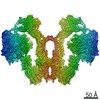
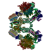
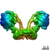
| Entry | Database: PDB / ID: 6hkt | ||||||
|---|---|---|---|---|---|---|---|
| Title | Structure of an H1-bound 6-nucleosome array | ||||||
 Components Components |
| ||||||
 Keywords Keywords | DNA BINDING PROTEIN / chromatin / nucleosome array / chromatin fiber | ||||||
| Function / homology |  Function and homology information Function and homology informationnegative regulation of tumor necrosis factor-mediated signaling pathway / negative regulation of megakaryocyte differentiation / protein localization to CENP-A containing chromatin / Chromatin modifying enzymes / Replacement of protamines by nucleosomes in the male pronucleus / CENP-A containing nucleosome / Packaging Of Telomere Ends / Negative Regulation of CDH1 Gene Transcription / Recognition and association of DNA glycosylase with site containing an affected purine / Cleavage of the damaged purine ...negative regulation of tumor necrosis factor-mediated signaling pathway / negative regulation of megakaryocyte differentiation / protein localization to CENP-A containing chromatin / Chromatin modifying enzymes / Replacement of protamines by nucleosomes in the male pronucleus / CENP-A containing nucleosome / Packaging Of Telomere Ends / Negative Regulation of CDH1 Gene Transcription / Recognition and association of DNA glycosylase with site containing an affected purine / Cleavage of the damaged purine / Deposition of new CENPA-containing nucleosomes at the centromere / telomere organization / Interleukin-7 signaling / Recognition and association of DNA glycosylase with site containing an affected pyrimidine / Cleavage of the damaged pyrimidine / RNA Polymerase I Promoter Opening / epigenetic regulation of gene expression / Inhibition of DNA recombination at telomere / Assembly of the ORC complex at the origin of replication / Meiotic synapsis / SUMOylation of chromatin organization proteins / Regulation of endogenous retroelements by the Human Silencing Hub (HUSH) complex / DNA methylation / Condensation of Prophase Chromosomes / Chromatin modifications during the maternal to zygotic transition (MZT) / SIRT1 negatively regulates rRNA expression / HCMV Late Events / ERCC6 (CSB) and EHMT2 (G9a) positively regulate rRNA expression / PRC2 methylates histones and DNA / Regulation of endogenous retroelements by KRAB-ZFP proteins / innate immune response in mucosa / Defective pyroptosis / HDACs deacetylate histones / Regulation of endogenous retroelements by Piwi-interacting RNAs (piRNAs) / RNA Polymerase I Promoter Escape / Nonhomologous End-Joining (NHEJ) / lipopolysaccharide binding / Transcriptional regulation by small RNAs / Formation of the beta-catenin:TCF transactivating complex / Activated PKN1 stimulates transcription of AR (androgen receptor) regulated genes KLK2 and KLK3 / RUNX1 regulates genes involved in megakaryocyte differentiation and platelet function / HDMs demethylate histones / G2/M DNA damage checkpoint / NoRC negatively regulates rRNA expression / B-WICH complex positively regulates rRNA expression / PKMTs methylate histone lysines / DNA Damage/Telomere Stress Induced Senescence / Pre-NOTCH Transcription and Translation / Meiotic recombination / Activation of anterior HOX genes in hindbrain development during early embryogenesis / Metalloprotease DUBs / Transcriptional regulation of granulopoiesis / RMTs methylate histone arginines / HCMV Early Events / antimicrobial humoral immune response mediated by antimicrobial peptide / structural constituent of chromatin / UCH proteinases / antibacterial humoral response / heterochromatin formation / nucleosome / nucleosome assembly / E3 ubiquitin ligases ubiquitinate target proteins / Recruitment and ATM-mediated phosphorylation of repair and signaling proteins at DNA double strand breaks / HATs acetylate histones / RUNX1 regulates transcription of genes involved in differentiation of HSCs / Factors involved in megakaryocyte development and platelet production / MLL4 and MLL3 complexes regulate expression of PPARG target genes in adipogenesis and hepatic steatosis / chromatin organization / Processing of DNA double-strand break ends / Senescence-Associated Secretory Phenotype (SASP) / Oxidative Stress Induced Senescence / defense response to Gram-negative bacterium / Estrogen-dependent gene expression / killing of cells of another organism / chromosome, telomeric region / defense response to Gram-positive bacterium / Ub-specific processing proteases / cadherin binding / Amyloid fiber formation / protein heterodimerization activity / negative regulation of cell population proliferation / protein-containing complex / extracellular space / DNA binding / RNA binding / extracellular exosome / extracellular region / nucleoplasm / nucleus / membrane / cytosol Similarity search - Function | ||||||
| Biological species |  Homo sapiens (human) Homo sapiens (human)synthetic construct (others) | ||||||
| Method |  X-RAY DIFFRACTION / SYNCHROTRON / MOLECULAR REPLACEMENT / Resolution: 9.7 Å X-RAY DIFFRACTION / SYNCHROTRON / MOLECULAR REPLACEMENT / Resolution: 9.7 Å | ||||||
 Authors Authors | Garcia-Saez, I. / Dimitrov, S. / Petosa, C. | ||||||
| Funding support |  France, 1items France, 1items
| ||||||
 Citation Citation | Journal: Mol. Cell / Year: 2018 Title: Structure of an H1-Bound 6-Nucleosome Array Reveals an Untwisted Two-Start Chromatin Fiber Conformation. Authors: Garcia-Saez, I. / Menoni, H. / Boopathi, R. / Shukla, M.S. / Soueidan, L. / Noirclerc-Savoye, M. / Le Roy, A. / Skoufias, D.A. / Bednar, J. / Hamiche, A. / Angelov, D. / Petosa, C. / Dimitrov, S. | ||||||
| History |
|
- Structure visualization
| Structure viewer | Molecule: MolmilJmol/JSmol |
|---|
- Downloads & links
-Download
| PDBx/mmCIF format | 6hkt.cif.gz | 1.8 MB | Display | PDBx/mmCIF format |
|---|---|---|---|---|
| PDB format | pdb6hkt.ent.gz | 1.4 MB | Display | PDB format |
| PDBx/mmJSON format | 6hkt.json.gz | Tree view | PDBx/mmJSON format | |
| Others |  Other downloads Other downloads |
-Validation report
| Arichive directory | https://data.pdbj.org/pub/pdb/validation_reports/hk/6hktftp://data.pdbj.org/pub/pdb/validation_reports/hk/6hkt | HTTPS FTP |
|---|
-Related structure data
| Related structure data |  3lz0S S: Starting model for refinement |
|---|---|
| Similar structure data |
-Links
 PDBj
PDBj


- Assembly
| Deposited unit | 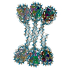
| ||||||||
|---|---|---|---|---|---|---|---|---|---|
| 1 |
| ||||||||
| Unit cell |
|
-Components
-Protein , 4 types, 48 molecules AEaeKOkoUYuyBFbfLPlpVZvzCGcgMQ...
| #1: Protein | Mass: 15719.445 Da / Num. of mol.: 12 Source method: isolated from a genetically manipulated source Details: THE 3 N-TERMINAL RESIDUES (GSH) COME FROM THE EXPRESSION TAG Source: (gene. exp.) Homo sapiens (human)Gene: HIST1H3A, H3FA, HIST1H3B, H3FL, HIST1H3C, H3FC, HIST1H3D, H3FB, HIST1H3E, H3FD, HIST1H3F, H3FI, HIST1H3G, H3FH, HIST1H3H, H3FK, HIST1H3I, H3FF, HIST1H3J, H3FJ Production host:  #2: Protein | Mass: 11676.703 Da / Num. of mol.: 12 Source method: isolated from a genetically manipulated source Details: THE 3 N-TERMINAL RESIDUES (GSH) COME FROM THE EXPRESSION TAG Source: (gene. exp.) Homo sapiens (human)Gene: HIST1H4A, H4/A, H4FA, HIST1H4B, H4/I, H4FI, HIST1H4C, H4/G, H4FG, HIST1H4D, H4/B, H4FB, HIST1H4E, H4/J, H4FJ, HIST1H4F, H4/C, H4FC, HIST1H4H, H4/H, H4FH, HIST1H4I, H4/M, H4FM, HIST1H4J, H4/E, ...Gene: HIST1H4A, H4/A, H4FA, HIST1H4B, H4/I, H4FI, HIST1H4C, H4/G, H4FG, HIST1H4D, H4/B, H4FB, HIST1H4E, H4/J, H4FJ, HIST1H4F, H4/C, H4FC, HIST1H4H, H4/H, H4FH, HIST1H4I, H4/M, H4FM, HIST1H4J, H4/E, H4FE, HIST1H4K, H4/D, H4FD, HIST1H4L, H4/K, H4FK, HIST2H4A, H4/N, H4F2, H4FN, HIST2H4, HIST2H4B, H4/O, H4FO, HIST4H4 Production host: #3: Protein | Mass: 14447.825 Da / Num. of mol.: 12 Source method: isolated from a genetically manipulated source Details: THE 3 N-TERMINAL RESIDUES (GSH) COME FROM THE EXPRESSION TAG Source: (gene. exp.) Homo sapiens (human) / Gene: HIST1H2AB, H2AFM, HIST1H2AE, H2AFA / Production host: #4: Protein | Mass: 14217.516 Da / Num. of mol.: 12 Source method: isolated from a genetically manipulated source Details: THE 3 N-TERMINAL RESIDUES (GSH) COME FROM THE EXPRESSION TAG Source: (gene. exp.) Homo sapiens (human) / Gene: HIST1H2BJ, H2BFR / Production host: |
|---|
-DNA chain , 2 types, 2 molecules IJ
| #5: DNA chain | Mass: 344755.281 Da / Num. of mol.: 1 / Source method: obtained synthetically Details: Six tandem 187-bp repeats containing Widom 601 sequence Source: (synth.) synthetic construct (others) |
|---|---|
| #6: DNA chain | Mass: 348512.312 Da / Num. of mol.: 1 / Source method: obtained synthetically Details: Six tandem 187-bp repeats containing Widom 601 sequence Source: (synth.) synthetic construct (others) |
-Experimental details
-Experiment
| Experiment | Method: X-RAY DIFFRACTION / Number of used crystals: 1 |
|---|
- Sample preparation
| Crystal | Density Matthews: 3.27 Å3/Da / Density % sol: 62.42 % |
|---|---|
| Crystal grow | Temperature: 293.15 K / Method: vapor diffusion, hanging drop / pH: 8.8 / Details: 15% MPD, 0.1 M NaCl and 0.1 M Tris pH 8.8 |
-Data collection
| Diffraction | Mean temperature: 100 K / Serial crystal experiment: N |
|---|---|
| Diffraction source | Source: SYNCHROTRON / Site: ESRF / Beamline: ID23-1 / Wavelength: 0.99187 Å |
| Detector | Type: DECTRIS PILATUS 6M-F / Detector: PIXEL / Date: Sep 11, 2015 |
| Radiation | Protocol: SINGLE WAVELENGTH / Monochromatic (M) / Laue (L): M / Scattering type: x-ray |
| Radiation wavelength | Wavelength: 0.99187 Å / Relative weight: 1 |
| Reflection | Resolution: 9.7→49.1 Å / Num. obs: 11252 / % possible obs: 99.2 % / Redundancy: 6.4 % / CC1/2: 0.998 / Rmerge(I) obs: 0.211 / Rpim(I) all: 0.09 / Rrim(I) all: 0.23 / Net I/σ(I): 7 |
| Reflection shell | Resolution: 9.7→10.85 Å / Redundancy: 6.8 % / Rmerge(I) obs: 0.881 / Mean I/σ(I) obs: 2.3 / Num. unique obs: 3152 / CC1/2: 0.604 / Rpim(I) all: 0.362 / % possible all: 100 |
- Processing
| Software |
| ||||||||||||||||||||||||||||||||||||||||||||||||||||||||
|---|---|---|---|---|---|---|---|---|---|---|---|---|---|---|---|---|---|---|---|---|---|---|---|---|---|---|---|---|---|---|---|---|---|---|---|---|---|---|---|---|---|---|---|---|---|---|---|---|---|---|---|---|---|---|---|---|---|
| Refinement | Method to determine structure: MOLECULAR REPLACEMENT Starting model: 3LZ0 Resolution: 9.7→49.074 Å / SU ML: 1.68 / Cross valid method: FREE R-VALUE / σ(F): 0.31 / Phase error: 34.02
| ||||||||||||||||||||||||||||||||||||||||||||||||||||||||
| Solvent computation | Shrinkage radii: 0.9 Å / VDW probe radii: 1.11 Å | ||||||||||||||||||||||||||||||||||||||||||||||||||||||||
| Refinement step | Cycle: LAST / Resolution: 9.7→49.074 Å
| ||||||||||||||||||||||||||||||||||||||||||||||||||||||||
| Refine LS restraints |
| ||||||||||||||||||||||||||||||||||||||||||||||||||||||||
| LS refinement shell |
|
