 Movie
Movie Controller

[English] 日本語
 Yorodumi
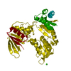
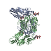
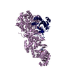
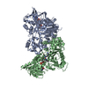
- PDB-6he2: Crystal structure of an open conformation of 2-Hydroxyisobutyryl-... -
+ Open data

- Basic information
| Entry | Database: PDB / ID: 6he2 | ||||||
|---|---|---|---|---|---|---|---|
| Title | Crystal structure of an open conformation of 2-Hydroxyisobutyryl-CoA Ligase (HCL) in complex with 2-HIB-AMP and CoA | ||||||
 Components Components | 2-hydroxyisobutyryl-CoA synthetase | ||||||
 Keywords Keywords | LIGASE / 2-hydroxyisobutyrate CoA | ||||||
| Function / homology |  Function and homology information Function and homology informationLigases; Forming carbon-sulfur bonds; Acid-thiol ligases / ligase activity / nucleotide binding Similarity search - Function | ||||||
| Biological species |  Aquincola tertiaricarbonis (bacteria) Aquincola tertiaricarbonis (bacteria) | ||||||
| Method |  X-RAY DIFFRACTION / SYNCHROTRON / MOLECULAR REPLACEMENT / Resolution: 2.3 Å X-RAY DIFFRACTION / SYNCHROTRON / MOLECULAR REPLACEMENT / Resolution: 2.3 Å | ||||||
 Authors Authors | Zahn, M. / Rohwerder, T. / Strater, N. | ||||||
 Citation Citation | Journal: J.Mol.Biol. / Year: 2019 Title: Structures of 2-Hydroxyisobutyric Acid-CoA Ligase Reveal Determinants of Substrate Specificity and Describe a Multi-Conformational Catalytic Cycle. Authors: Zahn, M. / Kurteva-Yaneva, N. / Schuster, J. / Krug, U. / Georgi, T. / Muller, R.H. / Rohwerder, T. / Strater, N. | ||||||
| History |
|
- Structure visualization
| Structure viewer | Molecule: MolmilJmol/JSmol |
|---|
- Downloads & links
-Download
| PDBx/mmCIF format | 6he2.cif.gz | 371 KB | Display | PDBx/mmCIF format |
|---|---|---|---|---|
| PDB format | pdb6he2.ent.gz | 299.2 KB | Display | PDB format |
| PDBx/mmJSON format | 6he2.json.gz | Tree view | PDBx/mmJSON format | |
| Others |  Other downloads Other downloads |
-Validation report
| Arichive directory | https://data.pdbj.org/pub/pdb/validation_reports/he/6he2ftp://data.pdbj.org/pub/pdb/validation_reports/he/6he2 | HTTPS FTP |
|---|
-Related structure data
| Related structure data | 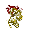 6hdwC 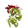 6hdxC 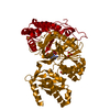 6hdyC  6he0C 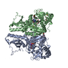 2y4oS S: Starting model for refinement C: citing same article ( |
|---|---|
| Similar structure data |

-Links
 PDBj
PDBj


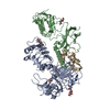
- Assembly
| Deposited unit | 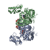
| ||||||||
|---|---|---|---|---|---|---|---|---|---|
| 1 |
| ||||||||
| Unit cell |
|
-Components
| #1: Protein | Mass: 55943.062 Da / Num. of mol.: 2 Source method: isolated from a genetically manipulated source Source: (gene. exp.) Aquincola tertiaricarbonis (bacteria) / Gene: hcl / Production host: References: UniProt: I3VE75, Ligases; Forming carbon-sulfur bonds; Acid-thiol ligases #2: Chemical |   Mass: 347.221 Da / Num. of mol.: 2 / Source method: obtained synthetically / Formula: C10H14N5O7P / Comment: AMP*YM Mass: 347.221 Da / Num. of mol.: 2 / Source method: obtained synthetically / Formula: C10H14N5O7P / Comment: AMP*YM#3: Chemical | 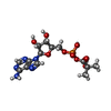  Mass: 433.310 Da / Num. of mol.: 2 / Source method: obtained synthetically / Formula: C14H20N5O9P Mass: 433.310 Da / Num. of mol.: 2 / Source method: obtained synthetically / Formula: C14H20N5O9P#4: Chemical |   Mass: 767.534 Da / Num. of mol.: 2 / Source method: obtained synthetically / Formula: C21H36N7O16P3S Mass: 767.534 Da / Num. of mol.: 2 / Source method: obtained synthetically / Formula: C21H36N7O16P3S#5: Water | ChemComp-HOH / |  Mass: 18.015 Da / Num. of mol.: 291 / Source method: isolated from a natural source / Formula: H2O Mass: 18.015 Da / Num. of mol.: 291 / Source method: isolated from a natural source / Formula: H2O |
|---|
-Experimental details
-Experiment
| Experiment | Method: X-RAY DIFFRACTION / Number of used crystals: 1 |
|---|
- Sample preparation
| Crystal | Density Matthews: 2.34 Å3/Da / Density % sol: 47.4 % |
|---|---|
| Crystal grow | Temperature: 293 K / Method: vapor diffusion, hanging drop Details: 0.1 M TRIS PH 8.0, 24% PEG 3350, VAPOR DIFFUSION, HANGING DROP, TEMPERATURE 293K |
-Data collection
| Diffraction | Mean temperature: 100 K |
|---|---|
| Diffraction source | Source: SYNCHROTRON / Site: BESSY  / Beamline: 14.1 / Wavelength: 0.918 Å / Beamline: 14.1 / Wavelength: 0.918 Å |
| Detector | Type: DECTRIS PILATUS3 6M / Detector: PIXEL / Date: Feb 8, 2013 |
| Radiation | Protocol: SINGLE WAVELENGTH / Monochromatic (M) / Laue (L): M / Scattering type: x-ray |
| Radiation wavelength | Wavelength: 0.918 Å / Relative weight: 1 |
| Reflection | Resolution: 2.3→40 Å / Num. obs: 47279 / % possible obs: 99.9 % / Redundancy: 5.6 % / Biso Wilson estimate: 45.74 Å2 / Rmerge(I) obs: 0.087 / Net I/σ(I): 11.6 |
| Reflection shell | Resolution: 2.3→2.42 Å / Redundancy: 5.7 % / Rmerge(I) obs: 0.472 / Mean I/σ(I) obs: 3.3 / Num. unique obs: 6830 / % possible all: 100 |
- Processing
| Software |
| |||||||||||||||||||||||||||||||||||||||||||||||||||||||||||||||||||||||||||||||||||||||||||||||||||||||||||||||||||||||||||||
|---|---|---|---|---|---|---|---|---|---|---|---|---|---|---|---|---|---|---|---|---|---|---|---|---|---|---|---|---|---|---|---|---|---|---|---|---|---|---|---|---|---|---|---|---|---|---|---|---|---|---|---|---|---|---|---|---|---|---|---|---|---|---|---|---|---|---|---|---|---|---|---|---|---|---|---|---|---|---|---|---|---|---|---|---|---|---|---|---|---|---|---|---|---|---|---|---|---|---|---|---|---|---|---|---|---|---|---|---|---|---|---|---|---|---|---|---|---|---|---|---|---|---|---|---|---|---|
| Refinement | Method to determine structure: MOLECULAR REPLACEMENT Starting model: 2Y4O Resolution: 2.3→39.72 Å / Cor.coef. Fo:Fc: 0.932 / Cor.coef. Fo:Fc free: 0.91 / SU R Cruickshank DPI: 0.279 / Cross valid method: THROUGHOUT / σ(F): 0 / SU R Blow DPI: 0.287 / SU Rfree Blow DPI: 0.203 / SU Rfree Cruickshank DPI: 0.203
| |||||||||||||||||||||||||||||||||||||||||||||||||||||||||||||||||||||||||||||||||||||||||||||||||||||||||||||||||||||||||||||
| Displacement parameters | Biso mean: 43.31 Å2
| |||||||||||||||||||||||||||||||||||||||||||||||||||||||||||||||||||||||||||||||||||||||||||||||||||||||||||||||||||||||||||||
| Refine analyze | Luzzati coordinate error obs: 0.27 Å | |||||||||||||||||||||||||||||||||||||||||||||||||||||||||||||||||||||||||||||||||||||||||||||||||||||||||||||||||||||||||||||
| Refinement step | Cycle: LAST / Resolution: 2.3→39.72 Å
| |||||||||||||||||||||||||||||||||||||||||||||||||||||||||||||||||||||||||||||||||||||||||||||||||||||||||||||||||||||||||||||
| Refine LS restraints |
| |||||||||||||||||||||||||||||||||||||||||||||||||||||||||||||||||||||||||||||||||||||||||||||||||||||||||||||||||||||||||||||
| LS refinement shell | Resolution: 2.3→2.36 Å / Total num. of bins used: 20
| |||||||||||||||||||||||||||||||||||||||||||||||||||||||||||||||||||||||||||||||||||||||||||||||||||||||||||||||||||||||||||||
| Refinement TLS params. | Method: refined / Refine-ID: X-RAY DIFFRACTION
| |||||||||||||||||||||||||||||||||||||||||||||||||||||||||||||||||||||||||||||||||||||||||||||||||||||||||||||||||||||||||||||
| Refinement TLS group |
|
