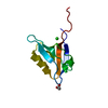
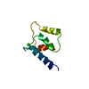
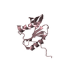
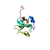
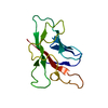
Entry Database : PDB / ID : 6h6rTitle Fragment Derived XIAP inhibitor E3 ubiquitin-protein ligase XIAP Keywords / / Function / homology Function Domain/homology Component
/ / / / / / / / / / / / / / / / / / / / / / / / / / / / / / / / / / / / / / / / / / / / / / / / / / / / / / / / / / / / / / / / / / / / / / / / / / / / / / / / / / / / Biological species Homo sapiens (human)Method / / Resolution : 2.03 Å Authors Williams, P.A. Journal : J. Med. Chem. / Year : 2018Title: A Fragment-Derived Clinical Candidate for Antagonism of X-Linked and Cellular Inhibitor of Apoptosis Proteins: 1-(6-[(4-Fluorophenyl)methyl]-5-(hydroxymethyl)-3,3-dimethyl-1 H,2 H,3 H- ... Title : A Fragment-Derived Clinical Candidate for Antagonism of X-Linked and Cellular Inhibitor of Apoptosis Proteins: 1-(6-[(4-Fluorophenyl)methyl]-5-(hydroxymethyl)-3,3-dimethyl-1 H,2 H,3 H-pyrrolo[3,2- b]pyridin-1-yl)-2-[(2 R,5 R)-5-methyl-2-([(3R)-3-methylmorpholin-4-yl]methyl)piperazin-1-yl]ethan-1-one (ASTX660).Authors: Johnson, C.N. / Ahn, J.S. / Buck, I.M. / Chiarparin, E. / Day, J.E.H. / Hopkins, A. / Howard, S. / Lewis, E.J. / Martins, V. / Millemaggi, A. / Munck, J.M. / Page, L.W. / Peakman, T. / ... Authors : Johnson, C.N. / Ahn, J.S. / Buck, I.M. / Chiarparin, E. / Day, J.E.H. / Hopkins, A. / Howard, S. / Lewis, E.J. / Martins, V. / Millemaggi, A. / Munck, J.M. / Page, L.W. / Peakman, T. / Reader, M. / Rich, S.J. / Saxty, G. / Smyth, T. / Thompson, N.T. / Ward, G.A. / Williams, P.A. / Wilsher, N.E. / Chessari, G. History Deposition Jul 30, 2018 Deposition site / Processing site Revision 1.0 Aug 22, 2018 Provider / Type Revision 1.1 Sep 5, 2018 Group / Database references / Category Item / _citation.page_first / _citation.page_lastRevision 1.2 May 15, 2024 Group / Database references / Derived calculationsCategory chem_comp_atom / chem_comp_bond ... chem_comp_atom / chem_comp_bond / database_2 / pdbx_struct_conn_angle / struct_conn Item _database_2.pdbx_DOI / _database_2.pdbx_database_accession ... _database_2.pdbx_DOI / _database_2.pdbx_database_accession / _pdbx_struct_conn_angle.ptnr1_auth_comp_id / _pdbx_struct_conn_angle.ptnr1_auth_seq_id / _pdbx_struct_conn_angle.ptnr1_label_asym_id / _pdbx_struct_conn_angle.ptnr1_label_comp_id / _pdbx_struct_conn_angle.ptnr1_label_seq_id / _pdbx_struct_conn_angle.ptnr1_symmetry / _pdbx_struct_conn_angle.ptnr3_auth_comp_id / _pdbx_struct_conn_angle.ptnr3_auth_seq_id / _pdbx_struct_conn_angle.ptnr3_label_asym_id / _pdbx_struct_conn_angle.ptnr3_label_comp_id / _pdbx_struct_conn_angle.ptnr3_label_seq_id / _pdbx_struct_conn_angle.ptnr3_symmetry / _pdbx_struct_conn_angle.value / _struct_conn.pdbx_dist_value / _struct_conn.ptnr1_auth_comp_id / _struct_conn.ptnr1_auth_seq_id / _struct_conn.ptnr1_label_asym_id / _struct_conn.ptnr1_label_atom_id / _struct_conn.ptnr1_label_comp_id / _struct_conn.ptnr1_label_seq_id / _struct_conn.ptnr2_auth_comp_id / _struct_conn.ptnr2_auth_seq_id / _struct_conn.ptnr2_label_asym_id / _struct_conn.ptnr2_label_atom_id / _struct_conn.ptnr2_label_comp_id / _struct_conn.ptnr2_symmetry
Show all Show less
 Movie
Movie Controller


 Open data
 Basic information
 Components
 Keywords
 Function and homology information
 Homo sapiens (human)
 X-RAY DIFFRACTION /
  Authors
 Citation
 Structure visualization
 Downloads & links
 Other downloads
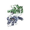


 PDBj
PDBj


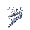
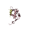
 Assembly
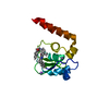


 Mass: 65.409 Da / Num. of mol.: 1 / Source method: obtained synthetically / Formula: Zn
 Mass: 22.990 Da / Num. of mol.: 1 / Source method: obtained synthetically / Formula: Na
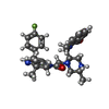
 Mass: 541.659 Da / Num. of mol.: 1 / Source method: obtained synthetically / Formula: C32H36FN5O2
 Mass: 18.015 Da / Num. of mol.: 197 / Source method: isolated from a natural source / Formula: H2O
 Sample preparation
 Processing