 Movie
Movie Controller

[English] 日本語
 Yorodumi
- PDB-6h5i: Single Particle Cryo-EM map of human Transferrin receptor 1 - H-F... -
+ Open data

- Basic information
| Entry | Database: PDB / ID: 6h5i | ||||||
|---|---|---|---|---|---|---|---|
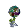
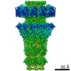
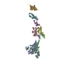
| Title | Single Particle Cryo-EM map of human Transferrin receptor 1 - H-Ferritin complex. | ||||||
 Components Components |
| ||||||
 Keywords Keywords | METAL BINDING PROTEIN / Transferrin Receptor 1 / Ferritin / Complex / Single Particle Cryo-EM | ||||||
| Function / homology |  Function and homology information Function and homology informationtransferrin receptor activity / postsynaptic recycling endosome membrane / negative regulation of mitochondrial fusion / positive regulation of isotype switching / Transferrin endocytosis and recycling / response to manganese ion / Differentiation of Keratinocytes in Interfollicular Epidermis in Mammalian Skin / iron ion sequestering activity / response to iron ion / RND1 GTPase cycle ...transferrin receptor activity / postsynaptic recycling endosome membrane / negative regulation of mitochondrial fusion / positive regulation of isotype switching / Transferrin endocytosis and recycling / response to manganese ion / Differentiation of Keratinocytes in Interfollicular Epidermis in Mammalian Skin / iron ion sequestering activity / response to iron ion / RND1 GTPase cycle / RND2 GTPase cycle / ferritin complex / Scavenging by Class A Receptors / RHOB GTPase cycle / response to copper ion / RHOJ GTPase cycle / RHOC GTPase cycle / Golgi Associated Vesicle Biogenesis / RHOQ GTPase cycle / ferroxidase / autolysosome / CDC42 GTPase cycle / negative regulation of ferroptosis / ferroxidase activity / RHOG GTPase cycle / RHOH GTPase cycle / RAC3 GTPase cycle / RHOA GTPase cycle / RAC2 GTPase cycle / response to retinoic acid / negative regulation of fibroblast proliferation / regulation of postsynaptic membrane neurotransmitter receptor levels / transport across blood-brain barrier / positive regulation of B cell proliferation / RAC1 GTPase cycle / clathrin-coated pit / osteoclast differentiation / response to nutrient / positive regulation of T cell proliferation / ferric iron binding / receptor-mediated endocytosis / autophagosome / Hsp70 protein binding / acute-phase response / iron ion transport / clathrin-coated endocytic vesicle membrane / HFE-transferrin receptor complex / transferrin transport / receptor internalization / positive regulation of protein-containing complex assembly / ferrous iron binding / Iron uptake and transport / cellular response to xenobiotic stimulus / positive regulation of protein localization to nucleus / multicellular organismal-level iron ion homeostasis / recycling endosome / recycling endosome membrane / tertiary granule lumen / melanosome / Cargo recognition for clathrin-mediated endocytosis / double-stranded RNA binding / Clathrin-mediated endocytosis / extracellular vesicle / virus receptor activity / cytoplasmic vesicle / blood microparticle / ficolin-1-rich granule lumen / basolateral plasma membrane / intracellular iron ion homeostasis / response to hypoxia / early endosome / positive regulation of canonical NF-kappaB signal transduction / cell surface receptor signaling pathway / lysosome / endosome / endosome membrane / intracellular signal transduction / immune response / iron ion binding / negative regulation of cell population proliferation / external side of plasma membrane / positive regulation of gene expression / Neutrophil degranulation / protein kinase binding / negative regulation of apoptotic process / protein-containing complex binding / perinuclear region of cytoplasm / glutamatergic synapse / cell surface / protein homodimerization activity / : / RNA binding / extracellular exosome / extracellular region / membrane / identical protein binding / nucleus / plasma membrane / cytosol / cytoplasm Similarity search - Function | ||||||
| Biological species |  Homo sapiens (human) Homo sapiens (human) | ||||||
| Method | ELECTRON MICROSCOPY / single particle reconstruction / cryo EM / Resolution: 3.9 Å | ||||||
 Authors Authors | Testi, C. / Montemiglio, L.C. / Vallone, B. / Des Georges, A. / Boffi, A. / Mancia, F. / Baiocco, P. / Savino, C. | ||||||
 Citation Citation | Journal: Nat Commun / Year: 2019 Title: Cryo-EM structure of the human ferritin-transferrin receptor 1 complex. Authors: Linda Celeste Montemiglio / Claudia Testi / Pierpaolo Ceci / Elisabetta Falvo / Martina Pitea / Carmelinda Savino / Alessandro Arcovito / Giovanna Peruzzi / Paola Baiocco / Filippo Mancia / ...Authors: Linda Celeste Montemiglio / Claudia Testi / Pierpaolo Ceci / Elisabetta Falvo / Martina Pitea / Carmelinda Savino / Alessandro Arcovito / Giovanna Peruzzi / Paola Baiocco / Filippo Mancia / Alberto Boffi / Amédée des Georges / Beatrice Vallone /   Abstract: Human transferrin receptor 1 (CD71) guarantees iron supply by endocytosis upon binding of iron-loaded transferrin and ferritin. Arenaviruses and the malaria parasite exploit CD71 for cell invasion ...Human transferrin receptor 1 (CD71) guarantees iron supply by endocytosis upon binding of iron-loaded transferrin and ferritin. Arenaviruses and the malaria parasite exploit CD71 for cell invasion and epitopes on CD71 for interaction with transferrin and pathogenic hosts were identified. Here, we provide the molecular basis of the CD71 ectodomain-human ferritin interaction by determining the 3.9 Å resolution single-particle cryo-electron microscopy structure of their complex and by validating our structural findings in a cellular context. The contact surfaces between the heavy-chain ferritin and CD71 largely overlap with arenaviruses and Plasmodium vivax binding regions in the apical part of the receptor ectodomain. Our data account for transferrin-independent binding of ferritin to CD71 and suggest that select pathogens may have adapted to enter cells by mimicking the ferritin access gate. | ||||||
| History |
|
- Structure visualization
| Movie |
Movie viewer |
|---|---|
| Structure viewer | Molecule: MolmilJmol/JSmol |
- Downloads & links
-Download
| PDBx/mmCIF format | 6h5i.cif.gz | 1009.8 KB | Display | PDBx/mmCIF format |
|---|---|---|---|---|
| PDB format | pdb6h5i.ent.gz | Display | PDB format | |
| PDBx/mmJSON format | 6h5i.json.gz | Tree view | PDBx/mmJSON format | |
| Others |  Other downloads Other downloads |
-Validation report
| Arichive directory | https://data.pdbj.org/pub/pdb/validation_reports/h5/6h5iftp://data.pdbj.org/pub/pdb/validation_reports/h5/6h5i | HTTPS FTP |
|---|
-Related structure data
| Related structure data |  0140MC  0046C  6gsrC M: map data used to model this data C: citing same article ( |
|---|---|
| Similar structure data |


-Links
 PDBj
PDBj


- Assembly
| Deposited unit | 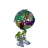
|
|---|---|
| 1 |
|
-Components
| #1: Protein | Mass: 20116.547 Da / Num. of mol.: 24 Source method: isolated from a genetically manipulated source Source: (gene. exp.) Homo sapiens (human) / Gene: FTH1, FTH, FTHL6, OK/SW-cl.84, PIG15 / Production host:  #2: Protein | Mass: 71807.258 Da / Num. of mol.: 2 Source method: isolated from a genetically manipulated source Source: (gene. exp.) Homo sapiens (human) / Gene: TFRC / Production host: Homo sapiens (human) / References: UniProt: P02786Has protein modification | Y | |
|---|
-Experimental details
-Experiment
| Experiment | Method: ELECTRON MICROSCOPY |
|---|---|
| EM experiment | Aggregation state: PARTICLE / 3D reconstruction method: single particle reconstruction |
- Sample preparation
| Component |
| ||||||||||||||||||||||||
|---|---|---|---|---|---|---|---|---|---|---|---|---|---|---|---|---|---|---|---|---|---|---|---|---|---|
| Source (natural) |
| ||||||||||||||||||||||||
| Source (recombinant) |
| ||||||||||||||||||||||||
| Buffer solution | pH: 7.2 | ||||||||||||||||||||||||
| Specimen | Conc.: 0.2 mg/ml / Embedding applied: NO / Shadowing applied: NO / Staining applied: NO / Vitrification applied: YES | ||||||||||||||||||||||||
| Vitrification | Cryogen name: ETHANE / Humidity: 100 % / Chamber temperature: 277 K |
- Electron microscopy imaging
| Experimental equipment |  Model: Titan Krios / Image courtesy: FEI Company |
|---|---|
| Microscopy | Model: FEI TITAN KRIOS |
| Electron gun | Electron source:  FIELD EMISSION GUN / Accelerating voltage: 300 kV / Illumination mode: OTHER FIELD EMISSION GUN / Accelerating voltage: 300 kV / Illumination mode: OTHER |
| Electron lens | Mode: OTHER |
| Image recording | Electron dose: 40 e/Å2 / Film or detector model: GATAN K2 SUMMIT (4k x 4k) |
- Processing
| Software | Name: PHENIX / Version: 1.13_2998: / Classification: refinement | ||||||||||||||||||||||||||||
|---|---|---|---|---|---|---|---|---|---|---|---|---|---|---|---|---|---|---|---|---|---|---|---|---|---|---|---|---|---|
| EM software |
| ||||||||||||||||||||||||||||
| CTF correction | Type: PHASE FLIPPING AND AMPLITUDE CORRECTION | ||||||||||||||||||||||||||||
| Particle selection | Num. of particles selected: 73700 | ||||||||||||||||||||||||||||
| Symmetry | Point symmetry: O (octahedral) | ||||||||||||||||||||||||||||
| 3D reconstruction | Resolution: 3.9 Å / Resolution method: FSC 0.143 CUT-OFF / Num. of particles: 53878 / Num. of class averages: 3 / Symmetry type: POINT | ||||||||||||||||||||||||||||
| Atomic model building | Protocol: OTHER | ||||||||||||||||||||||||||||
| Atomic model building |
| ||||||||||||||||||||||||||||
| Refine LS restraints |
|


