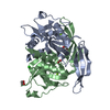
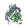
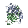
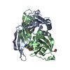
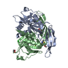
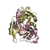
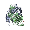
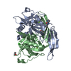
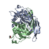
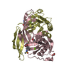
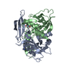
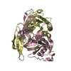
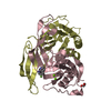
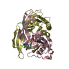
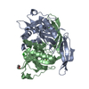
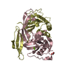
Entry Database : PDB / ID : 6gruTitle Crystal structure of human NUDT5 ADP-sugar pyrophosphatase Keywords / / / / Function / homology Function Domain/homology Component
/ / / / / / / / / / / / / / / / / / / / / / / / / / / / / / / / / / / / / / / / / Biological species Homo sapiens (human)Method / / / Resolution : 1.93 Å Authors Dubianok, Y. / Collins, P. / Krojer, T. / Fairhead, M. / MacLean, E. / Diaz Saez, L. / Strain-Damerell, C. / Elkins, J. / Burgess-Brown, N. / Bountra, C. ...Dubianok, Y. / Collins, P. / Krojer, T. / Fairhead, M. / MacLean, E. / Diaz Saez, L. / Strain-Damerell, C. / Elkins, J. / Burgess-Brown, N. / Bountra, C. / Arrowsmith, C.H. / Edwards, A. / Huber, K. / von Delft, F. / Structural Genomics Consortium (SGC) Journal : To Be Published Title : Crystal structure of human NUDT5Authors : Dubianok, Y. / Elkins, J. / Bountra, C. / Arrowsmith, C.H. / Edwards, A. / Huber, K. / von Delft, F. / Structural Genomics Consortium (SGC) History Deposition Jun 12, 2018 Deposition site / Processing site Revision 1.0 Jun 27, 2018 Provider / Type Revision 1.1 Jan 17, 2024 Group Data collection / Database references ... Data collection / Database references / Derived calculations / Refinement description Category chem_comp_atom / chem_comp_bond ... chem_comp_atom / chem_comp_bond / database_2 / pdbx_initial_refinement_model / pdbx_struct_conn_angle / struct_conn / struct_ncs_dom_lim Item _database_2.pdbx_DOI / _database_2.pdbx_database_accession ... _database_2.pdbx_DOI / _database_2.pdbx_database_accession / _pdbx_struct_conn_angle.ptnr1_auth_seq_id / _pdbx_struct_conn_angle.ptnr3_auth_seq_id / _pdbx_struct_conn_angle.value / _struct_conn.pdbx_dist_value / _struct_conn.ptnr1_auth_asym_id / _struct_conn.ptnr1_auth_comp_id / _struct_conn.ptnr1_auth_seq_id / _struct_conn.ptnr1_label_asym_id / _struct_conn.ptnr1_label_atom_id / _struct_conn.ptnr1_label_comp_id / _struct_conn.ptnr1_label_seq_id / _struct_conn.ptnr2_auth_asym_id / _struct_conn.ptnr2_auth_comp_id / _struct_conn.ptnr2_auth_seq_id / _struct_conn.ptnr2_label_asym_id / _struct_conn.ptnr2_label_atom_id / _struct_conn.ptnr2_label_comp_id / _struct_ncs_dom_lim.beg_auth_comp_id / _struct_ncs_dom_lim.beg_label_asym_id / _struct_ncs_dom_lim.beg_label_comp_id / _struct_ncs_dom_lim.beg_label_seq_id / _struct_ncs_dom_lim.end_auth_comp_id / _struct_ncs_dom_lim.end_label_asym_id / _struct_ncs_dom_lim.end_label_comp_id / _struct_ncs_dom_lim.end_label_seq_id
Show all Show less
 Movie
Movie Controller


 Open data
 Basic information
 Components
 Keywords
 Function and homology information
 Homo sapiens (human)
 X-RAY DIFFRACTION /
  Authors
 Citation
 Structure visualization
 Downloads & links
 Other downloads
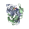

 PDBj
PDBj

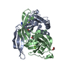
 Assembly


 Mass: 24.305 Da / Num. of mol.: 8 / Source method: obtained synthetically / Formula: Mg
 Mass: 35.453 Da / Num. of mol.: 1 / Source method: obtained synthetically / Formula: Cl
 Mass: 62.068 Da / Num. of mol.: 2 / Source method: obtained synthetically / Formula: C2H6O2
 Mass: 18.015 Da / Num. of mol.: 225 / Source method: isolated from a natural source / Formula: H2O
 Sample preparation
 / Beamline: I04-1 / Wavelength: 0.9159 Å
 Processing