 Movie
Movie Controller

[English] 日本語
 Yorodumi
- PDB-6gno: Crystal Structure Of Sea Bream Transthyretin in complex with Tetr... -
+ Open data

- Basic information
| Entry | Database: PDB / ID: 6gno | ||||||
|---|---|---|---|---|---|---|---|
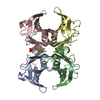
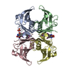
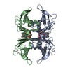
| Title | Crystal Structure Of Sea Bream Transthyretin in complex with Tetrabromobisphenol A (TBBPA) | ||||||
 Components Components | Transthyretin | ||||||
 Keywords Keywords | TRANSPORT PROTEIN / transthyretin / thyroxine disrupting chemicals / TDCs / Tetrabromobisphenol A (TBBPA) | ||||||
| Function / homology |  Function and homology information Function and homology informationthyroid hormone binding / purine nucleobase metabolic process / hormone activity / extracellular region Similarity search - Function | ||||||
| Biological species |  Sparus aurata (gilthead seabream) Sparus aurata (gilthead seabream) | ||||||
| Method |  X-RAY DIFFRACTION / MOLECULAR REPLACEMENT / Resolution: 1.65 Å X-RAY DIFFRACTION / MOLECULAR REPLACEMENT / Resolution: 1.65 Å | ||||||
 Authors Authors | Grundstrom, C. / Zhang, J. / Olofsson, A. / Andersson, P.L. / Sauer-Eriksson, A.E. | ||||||
| Funding support |  Sweden, 1items Sweden, 1items
| ||||||
 Citation Citation | Journal: Environ. Sci. Technol. / Year: 2018 Title: Interspecies Variation between Fish and Human Transthyretins in Their Binding of Thyroid-Disrupting Chemicals. Authors: Zhang, J. / Grundstrom, C. / Brannstrom, K. / Iakovleva, I. / Lindberg, M. / Olofsson, A. / Andersson, P.L. / Sauer-Eriksson, A.E. #1: Journal: Environ. Sci. Technol. / Year: 2016Title: Structure-Based Virtual Screening Protocol for in Silico Identification of Potential Thyroid Disrupting Chemicals Targeting Transthyretin. Authors: Zhang, J. / Begum, A. / Brannstrom, K. / Grundstrom, C. / Iakovleva, I. / Olofsson, A. / Sauer-Eriksson, A.E. / Andersson, P.L. | ||||||
| History |
|
- Structure visualization
| Structure viewer | Molecule: MolmilJmol/JSmol |
|---|
- Downloads & links
-Download
| PDBx/mmCIF format | 6gno.cif.gz | 114.8 KB | Display | PDBx/mmCIF format |
|---|---|---|---|---|
| PDB format | pdb6gno.ent.gz | 86.8 KB | Display | PDB format |
| PDBx/mmJSON format | 6gno.json.gz | Tree view | PDBx/mmJSON format | |
| Others |  Other downloads Other downloads |
-Validation report
| Arichive directory | https://data.pdbj.org/pub/pdb/validation_reports/gn/6gnoftp://data.pdbj.org/pub/pdb/validation_reports/gn/6gno | HTTPS FTP |
|---|
-Related structure data
| Related structure data | 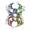 6gnmC  6gnpC  6gnrC  6gnwC  6gonC  6gooC  6gr7C  6grpC 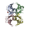 1sn2S S: Starting model for refinement C: citing same article ( |
|---|---|
| Similar structure data |


-Links
 PDBj
PDBj

- Assembly
| Deposited unit | 
| |||||||||
|---|---|---|---|---|---|---|---|---|---|---|
| 1 | 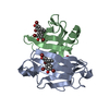
| |||||||||
| Unit cell |
| |||||||||
| Components on special symmetry positions |
|
-Components
| #1: Protein | Mass: 14455.096 Da / Num. of mol.: 2 Source method: isolated from a genetically manipulated source Source: (gene. exp.) Sparus aurata (gilthead seabream) / Production host:  #2: Chemical |   Mass: 543.871 Da / Num. of mol.: 2 / Source method: obtained synthetically / Formula: C15H12Br4O2 Mass: 543.871 Da / Num. of mol.: 2 / Source method: obtained synthetically / Formula: C15H12Br4O2#3: Chemical | ChemComp-SO4 / |   Mass: 96.063 Da / Num. of mol.: 1 / Source method: obtained synthetically / Formula: SO4 Mass: 96.063 Da / Num. of mol.: 1 / Source method: obtained synthetically / Formula: SO4#4: Water | ChemComp-HOH / |  Mass: 18.015 Da / Num. of mol.: 302 / Source method: isolated from a natural source / Formula: H2O Mass: 18.015 Da / Num. of mol.: 302 / Source method: isolated from a natural source / Formula: H2O |
|---|
-Experimental details
-Experiment
| Experiment | Method: X-RAY DIFFRACTION / Number of used crystals: 1 |
|---|
- Sample preparation
| Crystal | Density Matthews: 2.04 Å3/Da / Density % sol: 39.8 % |
|---|---|
| Crystal grow | Temperature: 291 K / Method: vapor diffusion, hanging drop / pH: 7.5 Details: Purified SaTTR in 100 mM NaCl and 20 mM Tris-HCl, pH 7.5, was concentrated to 5 mg per ml. TBBPA was added at 5 x molar excess to the protein. The reservoir contained 1.9 M AmSO4. Drop size 3 plus 3 microliter |
-Data collection
| Diffraction | Mean temperature: 100 K |
|---|---|
| Diffraction source | Source: ROTATING ANODE / Type: BRUKER AXS MICROSTAR-H / Wavelength: 1.5418 Å |
| Detector | Type: Bruker Platinum 135 / Detector: CCD / Date: Oct 18, 2016 |
| Radiation | Protocol: SINGLE WAVELENGTH / Monochromatic (M) / Laue (L): M / Scattering type: x-ray |
| Radiation wavelength | Wavelength: 1.5418 Å / Relative weight: 1 |
| Reflection | Resolution: 1.65→30.9 Å / Num. obs: 28420 / % possible obs: 97.2 % / Redundancy: 10.1 % / Rmerge(I) obs: 0.072 / Net I/σ(I): 29.7 |
| Reflection shell | Resolution: 1.65→1.71 Å / Redundancy: 2.9 % / Rmerge(I) obs: 0.426 / Mean I/σ(I) obs: 2.4 / Num. unique obs: 2599 / % possible all: 91 |
- Processing
| Software |
| |||||||||||||||||||||||||||||||||||||||||||||||||||||||||||||||||||||||||||||||||||||||||||||||||||||||||||||||||||||||||||||||||||||
|---|---|---|---|---|---|---|---|---|---|---|---|---|---|---|---|---|---|---|---|---|---|---|---|---|---|---|---|---|---|---|---|---|---|---|---|---|---|---|---|---|---|---|---|---|---|---|---|---|---|---|---|---|---|---|---|---|---|---|---|---|---|---|---|---|---|---|---|---|---|---|---|---|---|---|---|---|---|---|---|---|---|---|---|---|---|---|---|---|---|---|---|---|---|---|---|---|---|---|---|---|---|---|---|---|---|---|---|---|---|---|---|---|---|---|---|---|---|---|---|---|---|---|---|---|---|---|---|---|---|---|---|---|---|---|
| Refinement | Method to determine structure: MOLECULAR REPLACEMENT Starting model: 1sn2 Resolution: 1.65→30.895 Å / SU ML: 0.2 / Cross valid method: FREE R-VALUE / σ(F): 1.34 / Phase error: 25.9
| |||||||||||||||||||||||||||||||||||||||||||||||||||||||||||||||||||||||||||||||||||||||||||||||||||||||||||||||||||||||||||||||||||||
| Solvent computation | Shrinkage radii: 0.9 Å / VDW probe radii: 1.11 Å | |||||||||||||||||||||||||||||||||||||||||||||||||||||||||||||||||||||||||||||||||||||||||||||||||||||||||||||||||||||||||||||||||||||
| Refinement step | Cycle: LAST / Resolution: 1.65→30.895 Å
| |||||||||||||||||||||||||||||||||||||||||||||||||||||||||||||||||||||||||||||||||||||||||||||||||||||||||||||||||||||||||||||||||||||
| Refine LS restraints |
| |||||||||||||||||||||||||||||||||||||||||||||||||||||||||||||||||||||||||||||||||||||||||||||||||||||||||||||||||||||||||||||||||||||
| LS refinement shell |
|
