 Movie
Movie Controller


+ Open data

- Basic information
| Entry | Database: PDB / ID: 6g7z | ||||||
|---|---|---|---|---|---|---|---|
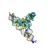
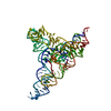
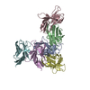
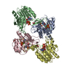
| Title | Lariat-capping ribozyme with a shortened DP2 stem loop | ||||||
 Components Components | (Lariat-capping ribozyme) x 2 | ||||||
 Keywords Keywords | RNA / Lariat capping ribozyme | ||||||
| Function / homology | RNA / RNA (> 10) / RNA (> 100) Function and homology information Function and homology information | ||||||
| Biological species |  Didymium iridis (eukaryote) Didymium iridis (eukaryote) | ||||||
| Method |  X-RAY DIFFRACTION / SYNCHROTRON / MOLECULAR REPLACEMENT / Resolution: 3.33594999571 Å X-RAY DIFFRACTION / SYNCHROTRON / MOLECULAR REPLACEMENT / Resolution: 3.33594999571 Å | ||||||
 Authors Authors | Masquida, B. / Meyer, M. / Olieric, V. | ||||||
 Citation Citation | Journal: Rna / Year: 2019 Title: Conformational adaptation of UNCG loops upon crowding. Authors: Meyer, M. / Walbott, H. / Olieric, V. / Kondo, J. / Costa, M. / Masquida, B. #1: Journal: Proc. Natl. Acad. Sci. U.S.A. / Year: 2014Title: Speciation of a group I intron into a lariat capping ribozyme. Authors: Meyer, M. / Nielsen, H. / Olieric, V. / Roblin, P. / Johansen, S.D. / Westhof, E. / Masquida, B. | ||||||
| History |
|
- Structure visualization
| Structure viewer | Molecule: MolmilJmol/JSmol |
|---|
- Downloads & links
-Download
| PDBx/mmCIF format | 6g7z.cif.gz | 253.5 KB | Display | PDBx/mmCIF format |
|---|---|---|---|---|
| PDB format | pdb6g7z.ent.gz | 170 KB | Display | PDB format |
| PDBx/mmJSON format | 6g7z.json.gz | Tree view | PDBx/mmJSON format | |
| Others |  Other downloads Other downloads |
-Validation report
| Arichive directory | https://data.pdbj.org/pub/pdb/validation_reports/g7/6g7zftp://data.pdbj.org/pub/pdb/validation_reports/g7/6g7z | HTTPS FTP |
|---|
-Related structure data
| Related structure data | 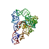 4p8zS S: Starting model for refinement |
|---|---|
| Similar structure data |


-Links
 PDBj
PDBj


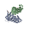
- Assembly
| Deposited unit | 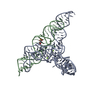
| ||||||||||||
|---|---|---|---|---|---|---|---|---|---|---|---|---|---|
| 1 |
| ||||||||||||
| Unit cell |
|
-Components
| #1: RNA chain | Mass: 42567.250 Da / Num. of mol.: 1 / Source method: obtained synthetically / Source: (synth.) Didymium iridis (eukaryote) | ||||
|---|---|---|---|---|---|
| #2: RNA chain | Mass: 17398.375 Da / Num. of mol.: 1 / Source method: obtained synthetically / Source: (synth.) Didymium iridis (eukaryote) | ||||
| #3: Chemical |   Mass: 24.305 Da / Num. of mol.: 2 / Source method: obtained synthetically / Formula: Mg Mass: 24.305 Da / Num. of mol.: 2 / Source method: obtained synthetically / Formula: Mg#4: Chemical | ChemComp-MES / |   Mass: 195.237 Da / Num. of mol.: 1 / Source method: obtained synthetically / Formula: C6H13NO4S / Comment: pH buffer*YM Mass: 195.237 Da / Num. of mol.: 1 / Source method: obtained synthetically / Formula: C6H13NO4S / Comment: pH buffer*YM#5: Water | ChemComp-HOH / |  Mass: 18.015 Da / Num. of mol.: 11 / Source method: isolated from a natural source / Formula: H2O Mass: 18.015 Da / Num. of mol.: 11 / Source method: isolated from a natural source / Formula: H2O |
-Experimental details
-Experiment
| Experiment | Method: X-RAY DIFFRACTION / Number of used crystals: 1 |
|---|
- Sample preparation
| Crystal | Density Matthews: 2.44 Å3/Da / Density % sol: 49.63 % / Description: rod-like 50 ?m long |
|---|---|
| Crystal grow | Temperature: 293 K / Method: vapor diffusion, sitting drop / pH: 7.5 / Details: 200 mM NaCl 100 mM HEPES pH 7.5 5-25% w/v PEG 3350 |
-Data collection
| Diffraction | Mean temperature: 100 K |
|---|---|
| Diffraction source | Source: SYNCHROTRON / Site: SLS  / Beamline: X06DA / Wavelength: 1 Å / Beamline: X06DA / Wavelength: 1 Å |
| Detector | Type: DECTRIS PILATUS 2M / Detector: PIXEL / Date: Sep 10, 2012 |
| Radiation | Protocol: SINGLE WAVELENGTH / Monochromatic (M) / Laue (L): M / Scattering type: x-ray |
| Radiation wavelength | Wavelength: 1 Å / Relative weight: 1 |
| Reflection | Resolution: 3.33→44.4 Å / Num. obs: 108094 / % possible obs: 94.1 % / Redundancy: 6.8 % / Biso Wilson estimate: 87.9280042935 Å2 / Net I/σ(I): 8.37 |
| Reflection shell | Resolution: 3.3359→3.8184 Å / Num. unique obs: 2173 |
- Processing
| Software |
| ||||||||||||||||||||||||||||||||||||||||
|---|---|---|---|---|---|---|---|---|---|---|---|---|---|---|---|---|---|---|---|---|---|---|---|---|---|---|---|---|---|---|---|---|---|---|---|---|---|---|---|---|---|
| Refinement | Method to determine structure: MOLECULAR REPLACEMENT Starting model: 4p8z Resolution: 3.33594999571→44.3955 Å / SU ML: 0.585939952286 / Cross valid method: FREE R-VALUE / σ(F): 2.00207155548 / Phase error: 41.5418750562
| ||||||||||||||||||||||||||||||||||||||||
| Solvent computation | Shrinkage radii: 0.9 Å / VDW probe radii: 1.11 Å | ||||||||||||||||||||||||||||||||||||||||
| Displacement parameters | Biso mean: 77.3605845159 Å2 | ||||||||||||||||||||||||||||||||||||||||
| Refinement step | Cycle: LAST / Resolution: 3.33594999571→44.3955 Å
| ||||||||||||||||||||||||||||||||||||||||
| Refine LS restraints |
| ||||||||||||||||||||||||||||||||||||||||
| LS refinement shell |
| ||||||||||||||||||||||||||||||||||||||||
| Refinement TLS params. | Method: refined / Origin x: 18.2824686099 Å / Origin y: -1.52786626811 Å / Origin z: 18.1738677075 Å
| ||||||||||||||||||||||||||||||||||||||||
| Refinement TLS group | Selection details: all |
