Mass: 18.015 Da / Num. of mol.: 229 / Source method: isolated from a natural source / Formula: H2O
-
Experimental details
-
Experiment
Experiment
Method: X-RAY DIFFRACTION / Number of used crystals: 1
-
Sample preparation
Crystal
Density Matthews: 2.32 Å3/Da / Density % sol: 47 % / Description: The crystals are small rods.
Crystal grow
Temperature: 293 K / Method: vapor diffusion, hanging drop / pH: 8.5 Details: The crystallisation plate was incluabed in 20 degree celcius for crystal growth. The reservoir had 500 micro litre of 20% PEG-MME 2000, 0.1M TRis (pH 8.5), 0.2M trimethylamine N-oxide. The ...Details: The crystallisation plate was incluabed in 20 degree celcius for crystal growth. The reservoir had 500 micro litre of 20% PEG-MME 2000, 0.1M TRis (pH 8.5), 0.2M trimethylamine N-oxide. The 2microlitre protein-peptide sample was mixed with 1ul of reservoir solution and placed in the cover slip for hanging drop set up
Method to determine structure: MOLECULAR REPLACEMENT / Resolution: 2.42→74.35 Å / Cor.coef. Fo:Fc: 0.954 / Cor.coef. Fo:Fc free: 0.911 / SU B: 14.457 / SU ML: 0.3 / Cross valid method: THROUGHOUT / ESU R: 0.552 / ESU R Free: 0.311 / Stereochemistry target values: MAXIMUM LIKELIHOOD / Details: HYDROGENS HAVE BEEN USED IF PRESENT IN THE INPUT
Rfactor
Num. reflection
% reflection
Selection details
Rfree
0.28035
1498
4.8 %
RANDOM
Rwork
0.21986
-
-
-
obs
0.22282
29906
98.16 %
-
Solvent computation
Ion probe radii: 0.8 Å / Shrinkage radii: 0.8 Å / VDW probe radii: 1.2 Å / Solvent model: MASK
 Movie
Movie Controller


 Open data
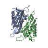
 Basic information
 Components
 Keywords
 Function and homology information
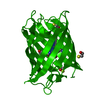
 Human respiratory syncytial virus A
 X-RAY DIFFRACTION /
  Authors
 United Kingdom, 1items
  Citation
 Structure visualization
 Downloads & links
 Other downloads


 PDBj
PDBj
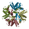
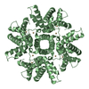
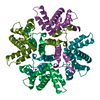
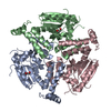
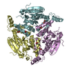
 Assembly
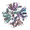


 Mass: 65.409 Da / Num. of mol.: 4 / Source method: obtained synthetically / Formula: Zn
 Mass: 18.015 Da / Num. of mol.: 229 / Source method: isolated from a natural source / Formula: H2O
 Sample preparation
 Processing