 Movie
Movie Controller

[English] 日本語
 Yorodumi
- PDB-6fv6: Monomer structure of the MATE family multidrug resistance transpo... -
+ Open data

- Basic information
| Entry | Database: PDB / ID: 6fv6 | ||||||||||||
|---|---|---|---|---|---|---|---|---|---|---|---|---|---|
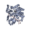
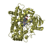
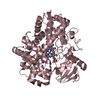
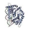
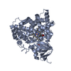
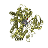
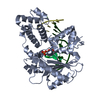
| Title | Monomer structure of the MATE family multidrug resistance transporter Aq_128 from Aquifex aeolicus in the outward-facing state | ||||||||||||
 Components Components | Aq128 | ||||||||||||
 Keywords Keywords | MEMBRANE PROTEIN / MATE class transporter | ||||||||||||
| Function / homology |  Function and homology information Function and homology informationFAD transmembrane transport / FMN transmembrane transporter activity / antiporter activity / xenobiotic transmembrane transporter activity / monoatomic ion transport / plasma membrane Similarity search - Function | ||||||||||||
| Biological species |   Aquifex aeolicus VF5 (bacteria) Aquifex aeolicus VF5 (bacteria) | ||||||||||||
| Method |  X-RAY DIFFRACTION / SYNCHROTRON / MOLECULAR REPLACEMENT / Resolution: 3.8 Å X-RAY DIFFRACTION / SYNCHROTRON / MOLECULAR REPLACEMENT / Resolution: 3.8 Å | ||||||||||||
 Authors Authors | Zhao, J. / Safarian, S. / Thielmann, Y. / Xie, H. / Wang, J. / Michel, H. | ||||||||||||
| Funding support |  Germany, Germany,  China, 3items China, 3items
| ||||||||||||
 Citation Citation | Journal: To Be Published Title: Monomer structure of Aq128 in the outward-facing state Authors: Zhao, J. / Safarian, S. / Thielmann, Y. / Xie, H. / Wang, J. / Michel, H. | ||||||||||||
| History |
|
- Structure visualization
| Structure viewer | Molecule: MolmilJmol/JSmol |
|---|
- Downloads & links
-Download
| PDBx/mmCIF format | 6fv6.cif.gz | 92 KB | Display | PDBx/mmCIF format |
|---|---|---|---|---|
| PDB format | pdb6fv6.ent.gz | 69.8 KB | Display | PDB format |
| PDBx/mmJSON format | 6fv6.json.gz | Tree view | PDBx/mmJSON format | |
| Others |  Other downloads Other downloads |
-Validation report
| Arichive directory | https://data.pdbj.org/pub/pdb/validation_reports/fv/6fv6ftp://data.pdbj.org/pub/pdb/validation_reports/fv/6fv6 | HTTPS FTP |
|---|
-Related structure data
| Related structure data | 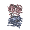 4mlbS S: Starting model for refinement |
|---|---|
| Similar structure data |


-Links
 PDBj
PDBj- Assembly
| Deposited unit | 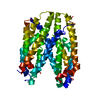
| ||||||||
|---|---|---|---|---|---|---|---|---|---|
| 1 |
| ||||||||
| Unit cell |
|
-Components
| #1: Protein | Mass: 52617.055 Da / Num. of mol.: 1 Source method: isolated from a genetically manipulated source Source: (gene. exp.) Aquifex aeolicus VF5 (bacteria) / Gene: aq_128 / Production host: |
|---|
-Experimental details
-Experiment
| Experiment | Method: X-RAY DIFFRACTION / Number of used crystals: 1 |
|---|
- Sample preparation
| Crystal | Density Matthews: 3.39 Å3/Da / Density % sol: 63.72 % / Description: plate |
|---|---|
| Crystal grow | Temperature: 291 K / Method: vapor diffusion / pH: 8.5 / Details: 0.1 M Tris 1.2 M ammonium sulphate |
-Data collection
| Diffraction | Mean temperature: 100 K |
|---|---|
| Diffraction source | Source: SYNCHROTRON / Site: SLS  / Beamline: X10SA / Wavelength: 1 Å / Beamline: X10SA / Wavelength: 1 Å |
| Detector | Type: PSI PILATUS 6M / Detector: PIXEL / Date: Mar 27, 2017 |
| Radiation | Protocol: SINGLE WAVELENGTH / Monochromatic (M) / Laue (L): M / Scattering type: x-ray |
| Radiation wavelength | Wavelength: 1 Å / Relative weight: 1 |
| Reflection | Resolution: 3.8→20 Å / Num. obs: 7452 / % possible obs: 99.9 % / Redundancy: 7.7 % / CC1/2: 0.999 / Rmerge(I) obs: 0.145 / Rpim(I) all: 0.057 / Rrim(I) all: 0.156 / Net I/σ(I): 9.6 |
| Reflection shell | Resolution: 3.8→3.94 Å / Redundancy: 8 % / Rmerge(I) obs: 1.778 / Mean I/σ(I) obs: 1.3 / Num. unique obs: 739 / CC1/2: 0.616 / Rpim(I) all: 0.664 / % possible all: 100 |
- Processing
| Software |
| ||||||||||||||||||||||||||||||||||||||||||
|---|---|---|---|---|---|---|---|---|---|---|---|---|---|---|---|---|---|---|---|---|---|---|---|---|---|---|---|---|---|---|---|---|---|---|---|---|---|---|---|---|---|---|---|
| Refinement | Method to determine structure: MOLECULAR REPLACEMENT Starting model: 4mlb Resolution: 3.8→19.972 Å / SU ML: 0.73 / Cross valid method: FREE R-VALUE / σ(F): 1.36 / Phase error: 40.63
| ||||||||||||||||||||||||||||||||||||||||||
| Solvent computation | Shrinkage radii: 0.9 Å / VDW probe radii: 1.11 Å | ||||||||||||||||||||||||||||||||||||||||||
| Refinement step | Cycle: LAST / Resolution: 3.8→19.972 Å
| ||||||||||||||||||||||||||||||||||||||||||
| Refine LS restraints |
| ||||||||||||||||||||||||||||||||||||||||||
| LS refinement shell |
|
