| Entry | Database: PDB / ID: 6fd3
|
|---|
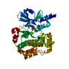
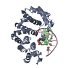
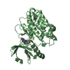
| Title | Thiophosphorylated PAK3 kinase domain |
|---|
 Components Components | Serine/threonine-protein kinase PAK 3 |
|---|
 Keywords Keywords | TRANSFERASE / kinase / thiophosphorylation / complex / ADP / phosphorylated |
|---|
| Function / homology |  Function and homology information Function and homology information
: / regulation of actin filament polymerization / dendritic spine morphogenesis / Activation of RAC1 / CD28 dependent Vav1 pathway / Ephrin signaling / regulation of postsynapse organization / regulation of axonogenesis / stimulatory C-type lectin receptor signaling pathway / RHOJ GTPase cycle ...: / regulation of actin filament polymerization / dendritic spine morphogenesis / Activation of RAC1 / CD28 dependent Vav1 pathway / Ephrin signaling / regulation of postsynapse organization / regulation of axonogenesis / stimulatory C-type lectin receptor signaling pathway / RHOJ GTPase cycle / MAP kinase kinase activity / RHOU GTPase cycle / dendrite development / RHO GTPases activate PAKs / CDC42 GTPase cycle / Generation of second messenger molecules / regulation of MAPK cascade / Sema3A PAK dependent Axon repulsion / ephrin receptor signaling pathway / RAC1 GTPase cycle / axonogenesis / CD209 (DC-SIGN) signaling / VEGFR2 mediated vascular permeability / regulation of actin cytoskeleton organization / SH3 domain binding / synapse organization / MAPK6/MAPK4 signaling / small GTPase binding / non-specific serine/threonine protein kinase / postsynaptic density / intracellular signal transduction / protein serine kinase activity / protein serine/threonine kinase activity / glutamatergic synapse / ATP binding / metal ion binding / plasma membrane / cytoplasm / cytosolSimilarity search - Function p21-activated kinase 3, catalytic domain / : / p21 activated kinase binding domain / CRIB domain superfamily / P21-Rho-binding domain / CRIB domain profile. / P21-Rho-binding domain / CRIB domain / Phosphorylase Kinase; domain 1 / Phosphorylase Kinase; domain 1 ...p21-activated kinase 3, catalytic domain / : / p21 activated kinase binding domain / CRIB domain superfamily / P21-Rho-binding domain / CRIB domain profile. / P21-Rho-binding domain / CRIB domain / Phosphorylase Kinase; domain 1 / Phosphorylase Kinase; domain 1 / Transferase(Phosphotransferase) domain 1 / Transferase(Phosphotransferase); domain 1 / Serine/threonine-protein kinase, active site / Serine/Threonine protein kinases active-site signature. / Protein kinase domain / Serine/Threonine protein kinases, catalytic domain / Protein kinase, ATP binding site / Protein kinases ATP-binding region signature. / Protein kinase domain profile. / Protein kinase domain / Protein kinase-like domain superfamily / 2-Layer Sandwich / Orthogonal Bundle / Mainly Alpha / Alpha BetaSimilarity search - Domain/homology |
|---|
| Biological species |  Homo sapiens (human) Homo sapiens (human) |
|---|
| Method |  X-RAY DIFFRACTION / SYNCHROTRON / MOLECULAR REPLACEMENT / molecular replacement / Resolution: 1.52 Å X-RAY DIFFRACTION / SYNCHROTRON / MOLECULAR REPLACEMENT / molecular replacement / Resolution: 1.52 Å |
|---|
 Authors Authors | Sorrell, F.J. / Wang, D. / von Delft, F. / Bountra, C. / Edwards, A.M. / Elkins, J.M. |
|---|
 Citation Citation | Journal: Biochem.J. / Year: 2019
Title: Solution structures and biophysical analysis of full-length group A PAKs reveal they are monomeric and auto-inhibited incis.
Authors: Sorrell, F.J. / Kilian, L.M. / Elkins, J.M. |
|---|
| History | | Deposition | Dec 21, 2017 | Deposition site: PDBE / Processing site: PDBE |
|---|
| Revision 1.0 | Jan 3, 2018 | Provider: repository / Type: Initial release |
|---|
| Revision 1.1 | Jul 17, 2019 | Group: Data collection / Database references / Category: citation / citation_author
Item: _citation.country / _citation.journal_abbrev ..._citation.country / _citation.journal_abbrev / _citation.journal_id_ASTM / _citation.journal_id_CSD / _citation.journal_id_ISSN / _citation.journal_volume / _citation.page_first / _citation.page_last / _citation.pdbx_database_id_DOI / _citation.pdbx_database_id_PubMed / _citation.title / _citation.year |
|---|
| Revision 1.2 | Jan 17, 2024 | Group: Data collection / Database references ...Data collection / Database references / Derived calculations / Refinement description
Category: chem_comp_atom / chem_comp_bond ...chem_comp_atom / chem_comp_bond / database_2 / pdbx_initial_refinement_model / pdbx_struct_conn_angle / struct_conn / struct_conn_type
Item: _database_2.pdbx_DOI / _database_2.pdbx_database_accession ..._database_2.pdbx_DOI / _database_2.pdbx_database_accession / _pdbx_struct_conn_angle.ptnr1_auth_comp_id / _pdbx_struct_conn_angle.ptnr1_auth_seq_id / _pdbx_struct_conn_angle.ptnr1_label_asym_id / _pdbx_struct_conn_angle.ptnr1_label_atom_id / _pdbx_struct_conn_angle.ptnr1_label_comp_id / _pdbx_struct_conn_angle.ptnr3_auth_comp_id / _pdbx_struct_conn_angle.ptnr3_auth_seq_id / _pdbx_struct_conn_angle.ptnr3_label_asym_id / _pdbx_struct_conn_angle.ptnr3_label_atom_id / _pdbx_struct_conn_angle.ptnr3_label_comp_id / _pdbx_struct_conn_angle.value / _struct_conn.conn_type_id / _struct_conn.id / _struct_conn.pdbx_dist_value / _struct_conn.pdbx_leaving_atom_flag / _struct_conn.ptnr1_auth_comp_id / _struct_conn.ptnr1_auth_seq_id / _struct_conn.ptnr1_label_atom_id / _struct_conn.ptnr1_label_comp_id / _struct_conn.ptnr1_label_seq_id / _struct_conn.ptnr2_auth_comp_id / _struct_conn.ptnr2_auth_seq_id / _struct_conn.ptnr2_label_asym_id / _struct_conn.ptnr2_label_atom_id / _struct_conn.ptnr2_label_comp_id / _struct_conn.ptnr2_label_seq_id / _struct_conn_type.id |
|---|
| Revision 1.3 | Nov 20, 2024 | Group: Structure summary / Category: pdbx_entry_details / pdbx_modification_feature |
|---|
|
|---|
 Movie
Movie Controller


 Open data
 Basic information
 Structure visualization
 Downloads & links
 Other downloads
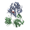


 PDBj
PDBj


 Assembly
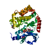


 Mass: 24.305 Da / Num. of mol.: 2 / Source method: obtained synthetically / Formula: Mg
 Mass: 427.201 Da / Num. of mol.: 1 / Source method: obtained synthetically / Formula: C10H15N5O10P2 / Comment: ADP, energy-carrying molecule*YM
 Mass: 62.068 Da / Num. of mol.: 2 / Source method: obtained synthetically / Formula: C2H6O2
 Mass: 18.015 Da / Num. of mol.: 276 / Source method: isolated from a natural source / Formula: H2O
 Sample preparation
 / Beamline: I03 / Wavelength: 0.976 Å
 Processing