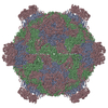
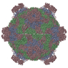
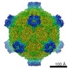
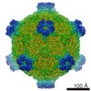
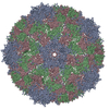
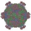
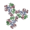
Journal: Curr Opin Virol / Year: 2020 Title: Virion structures and genome delivery of honeybee viruses. Authors: Michaela Procházková / Karel Škubník / Tibor Füzik / Liya Mukhamedova / Antonín Přidal / Pavel Plevka / Abstract: The western honeybee is the primary pollinator of numerous food crops. Furthermore, honeybees are essential for ecosystem stability by sustaining the diversity and abundance of wild flowering plants. ...The western honeybee is the primary pollinator of numerous food crops. Furthermore, honeybees are essential for ecosystem stability by sustaining the diversity and abundance of wild flowering plants. However, the worldwide population of honeybees is under pressure from environmental stress and pathogens. Viruses from the families Iflaviridae and Dicistroviridae, together with their vector, the parasitic mite Varroa destructor, are the major threat to the world's honeybees. Dicistroviruses and iflaviruses have capsids with icosahedral symmetries. Acidic pH triggers the genome release of both dicistroviruses and iflaviruses. The capsids of iflaviruses expand, whereas those of dicistroviruses remain compact until the genome release. Furthermore, dicistroviruses use inner capsid proteins, whereas iflaviruses employ protruding domains or minor capsid proteins from the virion surface to penetrate membranes and deliver their genomes into the cell cytoplasm. The structural characterization of the infection process opens up possibilities for the development of antiviral compounds.
History
Deposition
Dec 1, 2017
Deposition site: PDBE / Processing site: PDBE
Revision 1.0
Dec 12, 2018
Provider: repository / Type: Initial release
Revision 1.1
Jul 31, 2019
Group: Data collection / Refinement description / Category: refine
Average exposure time: 1 sec. / Electron dose: 21 e/Å2 / Detector mode: COUNTING / Film or detector model: FEI FALCON II (4k x 4k) / Num. of grids imaged: 1
Image scans
Width: 4096 / Height: 4096 / Movie frames/image: 16 / Used frames/image: 2-16
Resolution: 3.1 Å / Resolution method: FSC 0.143 CUT-OFF / Num. of particles: 21000 / Algorithm: FOURIER SPACE / Num. of class averages: 1 / Symmetry type: POINT
Atomic model building
Protocol: OTHER / Space: REAL
Refinement
Resolution: 3.1→243.399 Å / SU ML: 0.8 / σ(F): 0.04 / Phase error: 48.02 / Stereochemistry target values: ML
Rfactor
Num. reflection
% reflection
Rfree
0.3232
1996
0.21 %
Rwork
0.3232
-
-
obs
0.3232
956082
99.92 %
Solvent computation
Shrinkage radii: 0.9 Å / VDW probe radii: 1.11 Å / Solvent model: FLAT BULK SOLVENT MODEL
Refine LS restraints
Refine-ID
Type
Dev ideal
Number
ELECTRONMICROSCOPY
f_bond_d
0.006
36190
ELECTRONMICROSCOPY
f_angle_d
1.065
49365
ELECTRONMICROSCOPY
f_dihedral_angle_d
11.391
12910
ELECTRONMICROSCOPY
f_chiral_restr
0.097
5390
ELECTRONMICROSCOPY
f_plane_restr
0.007
6375
+
About Yorodumi
-
News
-
Feb 9, 2022. New format data for meta-information of EMDB entries
New format data for meta-information of EMDB entries
Version 3 of the EMDB header file is now the official format.
The previous official version 1.9 will be removed from the archive.
In the structure databanks used in Yorodumi, some data are registered as the other names, "COVID-19 virus" and "2019-nCoV". Here are the details of the virus and the list of structure data.
Jan 31, 2019. EMDB accession codes are about to change! (news from PDBe EMDB page)
EMDB accession codes are about to change! (news from PDBe EMDB page)
The allocation of 4 digits for EMDB accession codes will soon come to an end. Whilst these codes will remain in use, new EMDB accession codes will include an additional digit and will expand incrementally as the available range of codes is exhausted. The current 4-digit format prefixed with “EMD-” (i.e. EMD-XXXX) will advance to a 5-digit format (i.e. EMD-XXXXX), and so on. It is currently estimated that the 4-digit codes will be depleted around Spring 2019, at which point the 5-digit format will come into force.
The EM Navigator/Yorodumi systems omit the EMD- prefix.
Related info.:Q: What is EMD? / ID/Accession-code notation in Yorodumi/EM Navigator
Yorodumi is a browser for structure data from EMDB, PDB, SASBDB, etc.
This page is also the successor to EM Navigator detail page, and also detail information page/front-end page for Omokage search.
The word "yorodu" (or yorozu) is an old Japanese word meaning "ten thousand". "mi" (miru) is to see.
Related info.:EMDB / PDB / SASBDB / Comparison of 3 databanks / Yorodumi Search / Aug 31, 2016. New EM Navigator & Yorodumi / Yorodumi Papers / Jmol/JSmol / Function and homology information / Changes in new EM Navigator and Yorodumi
 Movie
Movie Controller


 Open data
 Basic information
 Components
 Keywords
 Function and homology information
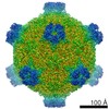 Deformed wing virus
 Authors
 Citation
 Structure visualization
 Downloads & links
 Other downloads
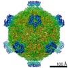

 PDBj
PDBj
 Assembly
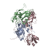


 Sample preparation
 Electron microscopy imaging
 FIELD EMISSION GUN / Accelerating voltage: 300 kV / Illumination mode: FLOOD BEAM
 Processing