 Movie
Movie Controller


+ Open data

- Basic information
| Entry | Database: PDB / ID: 6egc | ||||||
|---|---|---|---|---|---|---|---|
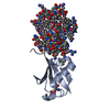
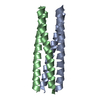
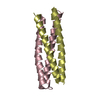
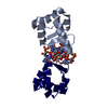
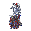
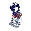
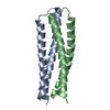
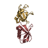
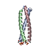
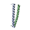
| Title | Single-chain version of 2L4HC2_23 (PDB 5J0K) | ||||||
 Components Components | SC_2L4HC2_23 | ||||||
 Keywords Keywords | DE NOVO PROTEIN / Computational Design / helical bundle / coiled-coil | ||||||
| Biological species | synthetic construct (others) | ||||||
| Method |  X-RAY DIFFRACTION / SYNCHROTRON / MOLECULAR REPLACEMENT / Resolution: 1.74 Å X-RAY DIFFRACTION / SYNCHROTRON / MOLECULAR REPLACEMENT / Resolution: 1.74 Å | ||||||
 Authors Authors | Bick, M.J. / Chen, Z. / DiMaio, F. | ||||||
| Funding support |  United States, 1items United States, 1items
| ||||||
 Citation Citation | Journal: J.Am.Chem.Soc. / Year: 2019 Title: Self-Assembling 2D Arrays with de Novo Protein Building Blocks. Authors: Chen, Z. / Johnson, M.C. / Chen, J. / Bick, M.J. / Boyken, S.E. / Lin, B. / De Yoreo, J.J. / Kollman, J.M. / Baker, D. / DiMaio, F. | ||||||
| History |
|
- Structure visualization
| Structure viewer | Molecule:  MolmilJmol/JSmol MolmilJmol/JSmol |
|---|
- Downloads & links
-Download
| PDBx/mmCIF format | 6egc.cif.gz | 95.4 KB | Display | PDBx/mmCIF format |
|---|---|---|---|---|
| PDB format | pdb6egc.ent.gz | 73.6 KB | Display | PDB format |
| PDBx/mmJSON format | 6egc.json.gz | Tree view | PDBx/mmJSON format | |
| Others |  Other downloads Other downloads |
-Validation report
| Arichive directory | https://data.pdbj.org/pub/pdb/validation_reports/eg/6egcftp://data.pdbj.org/pub/pdb/validation_reports/eg/6egc | HTTPS FTP |
|---|
-Related structure data
| Similar structure data |
|---|
-Links
 PDBj
PDBj
- Assembly
| Deposited unit | 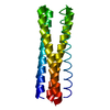
| ||||||||
|---|---|---|---|---|---|---|---|---|---|
| 1 |
| ||||||||
| Unit cell |
|
-Components
| #1: Protein | Mass: 19030.600 Da / Num. of mol.: 1 Source method: isolated from a genetically manipulated source Source: (gene. exp.) synthetic construct (others) / Production host:  |
|---|---|
| #2: Water | ChemComp-HOH /  Mass: 18.015 Da / Num. of mol.: 47 / Source method: isolated from a natural source / Formula: H2O Mass: 18.015 Da / Num. of mol.: 47 / Source method: isolated from a natural source / Formula: H2O |
-Experimental details
-Experiment
| Experiment | Method: X-RAY DIFFRACTION / Number of used crystals: 1 |
|---|
- Sample preparation
| Crystal | Density Matthews: 2.19 Å3/Da / Density % sol: 43.72 % |
|---|---|
| Crystal grow | Temperature: 291.15 K / Method: vapor diffusion, hanging drop / pH: 8.5 Details: Molecular Dimensions Morpheus H9 (0.1M Amino acids, 0.1M Buffer System 3 pH 8.5, 50% (v/v) Precipitant Mix 1) |
-Data collection
| Diffraction | Mean temperature: 100 K |
|---|---|
| Diffraction source | Source: SYNCHROTRON / Site: ALS / Beamline: 8.2.1 / Wavelength: 0.999978 Å |
| Detector | Type: ADSC QUANTUM 315r / Detector: CCD / Date: Mar 9, 2016 |
| Radiation | Monochromator: Double-crystal Si(111) and multilayer / Protocol: SINGLE WAVELENGTH / Monochromatic (M) / Laue (L): M / Scattering type: x-ray |
| Radiation wavelength | Wavelength: 0.999978 Å / Relative weight: 1 |
| Reflection | Resolution: 1.47→50 Å / Num. obs: 20818 / % possible obs: 75.5 % / Redundancy: 3.4 % / Biso Wilson estimate: 29 Å2 / Rmerge(I) obs: 0.055 / Rpim(I) all: 0.034 / Rrim(I) all: 0.064 / Χ2: 0.908 / Net I/av σ(I): 15.2 / Net I/σ(I): 7.3 |
| Reflection shell | Resolution: 1.47→1.5 Å / Redundancy: 1.1 % / Rmerge(I) obs: 2.723 / Mean I/σ(I) obs: 0.15 / Num. unique obs: 79 / CC1/2: 0.647 / Rrim(I) all: 3.794 / Χ2: 0.119 / % possible all: 5.8 |
- Processing
| Software |
| |||||||||||||||||||||||||||||||||||||||||||||||||||||||||||||||||||||||||||||||||||||||||||||||||||||||||||||||||||||||||||||
|---|---|---|---|---|---|---|---|---|---|---|---|---|---|---|---|---|---|---|---|---|---|---|---|---|---|---|---|---|---|---|---|---|---|---|---|---|---|---|---|---|---|---|---|---|---|---|---|---|---|---|---|---|---|---|---|---|---|---|---|---|---|---|---|---|---|---|---|---|---|---|---|---|---|---|---|---|---|---|---|---|---|---|---|---|---|---|---|---|---|---|---|---|---|---|---|---|---|---|---|---|---|---|---|---|---|---|---|---|---|---|---|---|---|---|---|---|---|---|---|---|---|---|---|---|---|---|
| Refinement | Method to determine structure: MOLECULAR REPLACEMENT Starting model: Computational design model Resolution: 1.74→22.52 Å / SU ML: 0.24 / Cross valid method: FREE R-VALUE / σ(F): 0 / Phase error: 32.84 Details: Iterative rounds of model building in Coot and refinement in Phenix.
| |||||||||||||||||||||||||||||||||||||||||||||||||||||||||||||||||||||||||||||||||||||||||||||||||||||||||||||||||||||||||||||
| Solvent computation | Shrinkage radii: 0.9 Å / VDW probe radii: 1.11 Å | |||||||||||||||||||||||||||||||||||||||||||||||||||||||||||||||||||||||||||||||||||||||||||||||||||||||||||||||||||||||||||||
| Displacement parameters | Biso mean: 58.9 Å2 | |||||||||||||||||||||||||||||||||||||||||||||||||||||||||||||||||||||||||||||||||||||||||||||||||||||||||||||||||||||||||||||
| Refinement step | Cycle: LAST / Resolution: 1.74→22.52 Å
| |||||||||||||||||||||||||||||||||||||||||||||||||||||||||||||||||||||||||||||||||||||||||||||||||||||||||||||||||||||||||||||
| Refine LS restraints |
| |||||||||||||||||||||||||||||||||||||||||||||||||||||||||||||||||||||||||||||||||||||||||||||||||||||||||||||||||||||||||||||
| LS refinement shell |
| |||||||||||||||||||||||||||||||||||||||||||||||||||||||||||||||||||||||||||||||||||||||||||||||||||||||||||||||||||||||||||||
| Refinement TLS params. | Method: refined / Refine-ID: X-RAY DIFFRACTION
| |||||||||||||||||||||||||||||||||||||||||||||||||||||||||||||||||||||||||||||||||||||||||||||||||||||||||||||||||||||||||||||
| Refinement TLS group |
|
