| Entry | Database: PDB / ID: 6eg7
|
|---|
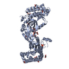
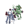
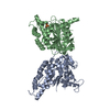
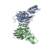
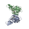
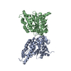
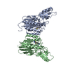
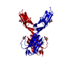
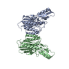
| Title | BbvCI B2 dimer with I3C clusters |
|---|
 Components Components | BbvCI endonuclease subunit 2 |
|---|
 Keywords Keywords | HYDROLASE / endonuclease / DNA binding protein / Type IIT restriction enzyme |
|---|
| Function / homology | Restriction endonuclease, type II, Bpu10I / Bpu10I restriction endonuclease / endonuclease activity / Chem-I3C / DI(HYDROXYETHYL)ETHER / BbvCI endonuclease subunit 2 Function and homology information Function and homology information |
|---|
| Biological species |  Brevibacillus brevis (bacteria) Brevibacillus brevis (bacteria) |
|---|
| Method |  X-RAY DIFFRACTION / SAD / Resolution: 3 Å X-RAY DIFFRACTION / SAD / Resolution: 3 Å |
|---|
 Authors Authors | Shen, B.W. / Stoddard, B.L. |
|---|
| Funding support |  United States, 1items United States, 1items | Organization | Grant number | Country |
|---|
| National Institutes of Health/National Human Genome Research Institute (NIH/NHGRI) | R01 GM105691 to BLS | United States |
|
|---|
 Citation Citation | Journal: Nucleic Acids Res. / Year: 2019
Title: Structure, subunit organization and behavior of the asymmetric Type IIT restriction endonuclease BbvCI.
Authors: Shen, B.W. / Doyle, L. / Bradley, P. / Heiter, D.F. / Lunnen, K.D. / Wilson, G.G. / Stoddard, B.L. |
|---|
| History | | Deposition | Aug 19, 2018 | Deposition site: RCSB / Processing site: RCSB |
|---|
| Revision 1.0 | Nov 14, 2018 | Provider: repository / Type: Initial release |
|---|
| Revision 1.1 | Nov 21, 2018 | Group: Data collection / Database references / Category: citation / citation_author
Item: _citation.pdbx_database_id_PubMed / _citation.title / _citation_author.name |
|---|
| Revision 1.2 | Jan 23, 2019 | Group: Data collection / Database references / Category: citation
Item: _citation.journal_volume / _citation.page_first ..._citation.journal_volume / _citation.page_first / _citation.page_last / _citation.year |
|---|
| Revision 1.3 | Dec 18, 2019 | Group: Author supporting evidence / Category: pdbx_audit_support / Item: _pdbx_audit_support.funding_organization |
|---|
| Revision 1.4 | Mar 13, 2024 | Group: Data collection / Database references / Refinement description
Category: chem_comp_atom / chem_comp_bond ...chem_comp_atom / chem_comp_bond / database_2 / struct_ncs_dom_lim
Item: _database_2.pdbx_DOI / _database_2.pdbx_database_accession ..._database_2.pdbx_DOI / _database_2.pdbx_database_accession / _struct_ncs_dom_lim.beg_auth_comp_id / _struct_ncs_dom_lim.beg_label_asym_id / _struct_ncs_dom_lim.beg_label_comp_id / _struct_ncs_dom_lim.beg_label_seq_id / _struct_ncs_dom_lim.end_auth_comp_id / _struct_ncs_dom_lim.end_label_asym_id / _struct_ncs_dom_lim.end_label_comp_id / _struct_ncs_dom_lim.end_label_seq_id |
|---|
|
|---|
 Movie
Movie Controller


 Open data
 Basic information
 Structure visualization
 Downloads & links
 Other downloads

 PDBj
PDBj Assembly
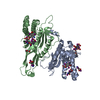

 Mass: 62.068 Da / Num. of mol.: 3 / Source method: obtained synthetically / Formula: C2H6O2
 Mass: 106.120 Da / Num. of mol.: 2 / Source method: isolated from a natural source / Formula: C4H10O3
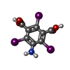
 Mass: 558.835 Da / Num. of mol.: 8 / Source method: obtained synthetically / Formula: C8H4I3NO4
 Mass: 18.015 Da / Num. of mol.: 9 / Source method: isolated from a natural source / Formula: H2O
 Sample preparation
 Processing