 Movie
Movie Controller

[English] 日本語
 Yorodumi
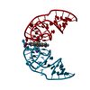
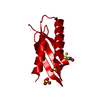
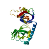
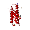
- PDB-6e82: Crystal structure of the Corn aptamer mutant A14U in complex with ThT -
+ Open data

- Basic information
| Entry | Database: PDB / ID: 6.0E+82 | ||||||
|---|---|---|---|---|---|---|---|
| Title | Crystal structure of the Corn aptamer mutant A14U in complex with ThT | ||||||
 Components Components | RNA (36-MER) | ||||||
 Keywords Keywords | RNA / polyribonucleotide / fluorigenic aptamer / G-quadruplex / thioflavin T / Corn mutant A14U | ||||||
| Function / homology | : / Chem-TFX / RNA / RNA (> 10) Function and homology information Function and homology information | ||||||
| Biological species | synthetic construct (others) | ||||||
| Method |  X-RAY DIFFRACTION / SYNCHROTRON / MOLECULAR REPLACEMENT / molecular replacement / Resolution: 3.101 Å X-RAY DIFFRACTION / SYNCHROTRON / MOLECULAR REPLACEMENT / molecular replacement / Resolution: 3.101 Å | ||||||
 Authors Authors | Sjekloca, L. / Ferre-D'Amare, A.R. | ||||||
 Citation Citation | Journal: Cell Chem Biol / Year: 2019 Title: Binding between G Quadruplexes at the Homodimer Interface of the Corn RNA Aptamer Strongly Activates Thioflavin T Fluorescence. Authors: Sjekloca, L. / Ferre-D'Amare, A.R. | ||||||
| History |
|
- Structure visualization
| Structure viewer | Molecule: MolmilJmol/JSmol |
|---|
- Downloads & links
-Download
| PDBx/mmCIF format | 6e82.cif.gz | 54.9 KB | Display | PDBx/mmCIF format |
|---|---|---|---|---|
| PDB format | pdb6e82.ent.gz | 40.6 KB | Display | PDB format |
| PDBx/mmJSON format | 6e82.json.gz | Tree view | PDBx/mmJSON format | |
| Others |  Other downloads Other downloads |
-Validation report
| Arichive directory | https://data.pdbj.org/pub/pdb/validation_reports/e8/6e82ftp://data.pdbj.org/pub/pdb/validation_reports/e8/6e82 | HTTPS FTP |
|---|
-Related structure data
| Related structure data |  6e80C  6e81C  6e84C  5bjpS C: citing same article ( S: Starting model for refinement |
|---|---|
| Similar structure data |

-Links
 PDBj
PDBj


- Assembly
| Deposited unit | 
| ||||||||
|---|---|---|---|---|---|---|---|---|---|
| 1 | 
| ||||||||
| Unit cell |
| ||||||||
| Components on special symmetry positions |
|
-Components
| #1: RNA chain | Mass: 11771.060 Da / Num. of mol.: 1 / Mutation: A14U / Source method: obtained synthetically / Source: (synth.) synthetic construct (others) |
|---|---|
| #2: Chemical | ChemComp-K /   Mass: 39.098 Da / Num. of mol.: 1 / Source method: obtained synthetically / Formula: K Mass: 39.098 Da / Num. of mol.: 1 / Source method: obtained synthetically / Formula: K |
| #3: Chemical | ChemComp-TFX /   Mass: 283.411 Da / Num. of mol.: 1 / Source method: obtained synthetically / Formula: C17H19N2S Mass: 283.411 Da / Num. of mol.: 1 / Source method: obtained synthetically / Formula: C17H19N2S |
-Experimental details
-Experiment
| Experiment | Method: X-RAY DIFFRACTION / Number of used crystals: 1 |
|---|
- Sample preparation
| Crystal | Density Matthews: 2.84 Å3/Da / Density % sol: 56.7 % / Description: rectangular plate-shaped |
|---|---|
| Crystal grow | Temperature: 294 K / Method: vapor diffusion, hanging drop Details: 20 % PEG 4000, 5% PEG 400, 10% glycerol, 0.2 M ammonium acetate pH 6.7, 0.1 M sodium citrate pH 5.6, 0.5 mM ThT |
-Data collection
| Diffraction | Mean temperature: 100 K | ||||||||||||||||||||||||
|---|---|---|---|---|---|---|---|---|---|---|---|---|---|---|---|---|---|---|---|---|---|---|---|---|---|
| Diffraction source | Source: SYNCHROTRON / Site: ALS  / Beamline: 5.0.1 / Wavelength: 1 Å / Beamline: 5.0.1 / Wavelength: 1 Å | ||||||||||||||||||||||||
| Detector | Type: DECTRIS PILATUS3 6M / Detector: PIXEL / Date: May 3, 2018 | ||||||||||||||||||||||||
| Radiation | Protocol: SINGLE WAVELENGTH / Monochromatic (M) / Laue (L): M / Scattering type: x-ray | ||||||||||||||||||||||||
| Radiation wavelength | Wavelength: 1 Å / Relative weight: 1 | ||||||||||||||||||||||||
| Reflection | Resolution: 2.96→37.74 Å / Num. obs: 2840 / % possible obs: 99.9 % / Redundancy: 19 % / Biso Wilson estimate: 75.66 Å2 / CC1/2: 1 / Rmerge(I) obs: 0.137 / Rpim(I) all: 0.032 / Rrim(I) all: 0.141 / Net I/σ(I): 13.8 / Num. measured all: 53903 / Scaling rejects: 45 | ||||||||||||||||||||||||
| Reflection shell | Diffraction-ID: 1
|
-Phasing
| Phasing | Method: molecular replacement | |||||||||
|---|---|---|---|---|---|---|---|---|---|---|
| Phasing MR |
|
- Processing
| Software |
| ||||||||||||||||||||||||||||||||||||||||
|---|---|---|---|---|---|---|---|---|---|---|---|---|---|---|---|---|---|---|---|---|---|---|---|---|---|---|---|---|---|---|---|---|---|---|---|---|---|---|---|---|---|
| Refinement | Method to determine structure: MOLECULAR REPLACEMENT Starting model: 5BJP Resolution: 3.101→33.031 Å / SU ML: 0.35 / Cross valid method: THROUGHOUT / σ(F): 1.59 / Phase error: 24.76
| ||||||||||||||||||||||||||||||||||||||||
| Solvent computation | Shrinkage radii: 0.9 Å / VDW probe radii: 1.11 Å | ||||||||||||||||||||||||||||||||||||||||
| Displacement parameters | Biso max: 184.27 Å2 / Biso mean: 108.9614 Å2 / Biso min: 63.98 Å2 | ||||||||||||||||||||||||||||||||||||||||
| Refinement step | Cycle: final / Resolution: 3.101→33.031 Å
| ||||||||||||||||||||||||||||||||||||||||
| Refine LS restraints |
| ||||||||||||||||||||||||||||||||||||||||
| LS refinement shell | Refine-ID: X-RAY DIFFRACTION / Rfactor Rfree error: 0 / Total num. of bins used: 2
| ||||||||||||||||||||||||||||||||||||||||
| Refinement TLS params. | Method: refined / Origin x: 1.8666 Å / Origin y: 50.7071 Å / Origin z: 20.137 Å
| ||||||||||||||||||||||||||||||||||||||||
| Refinement TLS group |
|
