 Movie
Movie Controller


+ Open data

- Basic information
| Entry | Database: PDB / ID: 6dz1 | ||||||
|---|---|---|---|---|---|---|---|
| Title | Studies of Ion Transport in K+ Channels | ||||||
 Components Components | Potassium channel protein | ||||||
 Keywords Keywords | MEMBRANE PROTEIN / K ion transport channel | ||||||
| Function / homology | Helix Hairpins - #70 / Potassium channel domain / Ion channel / membrane => GO:0016020 / Helix Hairpins / Orthogonal Bundle / Mainly Alpha / : / Potassium channel protein Function and homology information Function and homology information | ||||||
| Biological species |  | ||||||
| Method |  X-RAY DIFFRACTION / SYNCHROTRON / SAD / Resolution: 2.26 Å X-RAY DIFFRACTION / SYNCHROTRON / SAD / Resolution: 2.26 Å | ||||||
 Authors Authors | Langan, P.S. / Vandavasi, V.G. / Weiss, K.L. / Wagner, A. / Duman, R. / El Omari, K. / Afonine, P.V. / Coates, L. | ||||||
 Citation Citation | Journal: Nat Commun / Year: 2018 Title: Anomalous X-ray diffraction studies of ion transport in K+channels. Authors: Langan, P.S. / Vandavasi, V.G. / Weiss, K.L. / Afonine, P.V. / El Omari, K. / Duman, R. / Wagner, A. / Coates, L. | ||||||
| History |
|
- Structure visualization
| Structure viewer | Molecule: MolmilJmol/JSmol |
|---|
- Downloads & links
-Download
| PDBx/mmCIF format | 6dz1.cif.gz | 52.1 KB | Display | PDBx/mmCIF format |
|---|---|---|---|---|
| PDB format | pdb6dz1.ent.gz | 36.7 KB | Display | PDB format |
| PDBx/mmJSON format | 6dz1.json.gz | Tree view | PDBx/mmJSON format | |
| Others |  Other downloads Other downloads |
-Validation report
| Arichive directory | https://data.pdbj.org/pub/pdb/validation_reports/dz/6dz1ftp://data.pdbj.org/pub/pdb/validation_reports/dz/6dz1 | HTTPS FTP |
|---|
-Related structure data
| Similar structure data |
|---|

-Links
 PDBj
PDBj

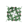
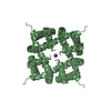
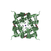
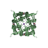
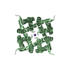
- Assembly
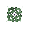
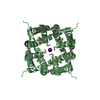
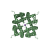
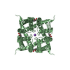
| Deposited unit | 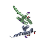
| ||||||||||||||||||||||||||||||
|---|---|---|---|---|---|---|---|---|---|---|---|---|---|---|---|---|---|---|---|---|---|---|---|---|---|---|---|---|---|---|---|
| 1 | 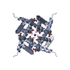
| ||||||||||||||||||||||||||||||
| 2 | 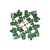
| ||||||||||||||||||||||||||||||
| Unit cell |
| ||||||||||||||||||||||||||||||
| Components on special symmetry positions |
|
-Components
| #1: Protein | Mass: 10569.399 Da / Num. of mol.: 2 Source method: isolated from a genetically manipulated source Source: (gene. exp.) #2: Chemical | ChemComp-K /   Mass: 39.098 Da / Num. of mol.: 10 Mass: 39.098 Da / Num. of mol.: 10Source method: isolated from a genetically manipulated source Formula: K #3: Chemical |   Mass: 118.174 Da / Num. of mol.: 3 / Source method: obtained synthetically / Formula: C6H14O2 / Comment: precipitant*YM Mass: 118.174 Da / Num. of mol.: 3 / Source method: obtained synthetically / Formula: C6H14O2 / Comment: precipitant*YM#4: Water | ChemComp-HOH / |  Mass: 18.015 Da / Num. of mol.: 26 / Source method: isolated from a natural source / Formula: H2O Mass: 18.015 Da / Num. of mol.: 26 / Source method: isolated from a natural source / Formula: H2O |
|---|
-Experimental details
-Experiment
| Experiment | Method: X-RAY DIFFRACTION / Number of used crystals: 1 |
|---|
- Sample preparation
| Crystal | Density Matthews: 2.45 Å3/Da / Density % sol: 49.77 % |
|---|---|
| Crystal grow | Temperature: 293 K / Method: vapor diffusion Details: Prepared by mixing equal volumes of protein solution in buffer (20mM Tris:HCl pH 7.8, 100mM KCl, and 4mM Anagrade DM) with well solution (72.5%MPD, 100mM KCl, and 100mM MES pH 6) |
-Data collection
| Diffraction | Mean temperature: 100 K |
|---|---|
| Diffraction source | Source: SYNCHROTRON / Site: Diamond  / Beamline: I23 / Wavelength: 3.35 Å / Beamline: I23 / Wavelength: 3.35 Å |
| Detector | Type: DECTRIS PILATUS 300K / Detector: PIXEL / Date: Jul 1, 2017 |
| Radiation | Protocol: SINGLE WAVELENGTH / Monochromatic (M) / Laue (L): M / Scattering type: x-ray |
| Radiation wavelength | Wavelength: 3.35 Å / Relative weight: 1 |
| Reflection | Resolution: 2.26→54.15 Å / Num. obs: 18417 / % possible obs: 97.8 % / Redundancy: 8.2 % / CC1/2: 0.994 / Rmerge(I) obs: 0.037 / Rpim(I) all: 0.019 / Net I/σ(I): 33.4 |
| Reflection shell | Resolution: 2.26→2.33 Å / Redundancy: 6.3 % / Rmerge(I) obs: 0.129 / Mean I/σ(I) obs: 10.7 / CC1/2: 0.987 / Rpim(I) all: 0.082 / % possible all: 93.4 |
- Processing
| Software |
| ||||||||||||||||||||||||||||||||||||||||||||||||||||||||
|---|---|---|---|---|---|---|---|---|---|---|---|---|---|---|---|---|---|---|---|---|---|---|---|---|---|---|---|---|---|---|---|---|---|---|---|---|---|---|---|---|---|---|---|---|---|---|---|---|---|---|---|---|---|---|---|---|---|
| Refinement | Method to determine structure: SAD / Resolution: 2.26→54.154 Å / SU ML: 0.21 / Cross valid method: FREE R-VALUE / σ(F): 1.58 / Phase error: 21.61 / Stereochemistry target values: ML
| ||||||||||||||||||||||||||||||||||||||||||||||||||||||||
| Solvent computation | Shrinkage radii: 0.9 Å / VDW probe radii: 1.11 Å / Solvent model: FLAT BULK SOLVENT MODEL | ||||||||||||||||||||||||||||||||||||||||||||||||||||||||
| Refinement step | Cycle: LAST / Resolution: 2.26→54.154 Å
| ||||||||||||||||||||||||||||||||||||||||||||||||||||||||
| Refine LS restraints |
| ||||||||||||||||||||||||||||||||||||||||||||||||||||||||
| LS refinement shell |
|
